

doi: 10.1212/WNL.0000000000003735.
Epub 2017 Feb 15.
Distribution map of gadolinium deposition within the cerebellum following GBCA administration
Affiliations
- PMID: 28202695
- PMCID: PMC11279554
- DOI: 10.1212/WNL.0000000000003735
Item in Clipboard
Distribution map of gadolinium deposition within the cerebellum following GBCA administration
Neurology.
.
No abstract available
Figures
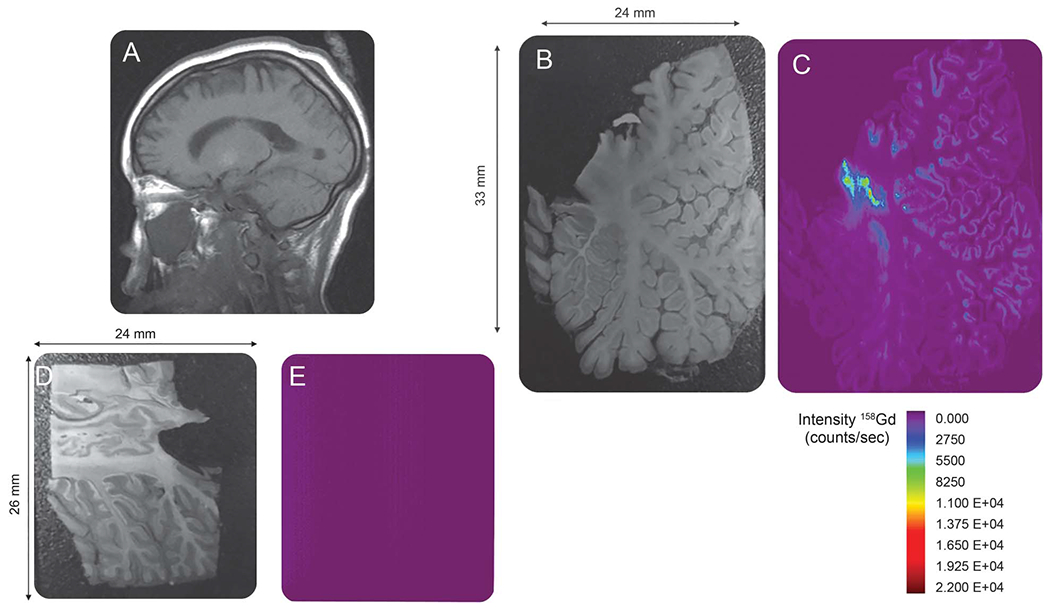
(A) Sagittal image through the level of the dentate nucleus prior to the last dose of gadolinium-based contrast agent (GBCA). No hyperintense signal is demonstrated within the dentate nucleus. (Note that the globus pallidus demonstrates T1 shortening due to hypoxic ischemic injury.) (B) Autopsy section through the cerebellum. (C) Laser ablation inductively coupled plasma mass spectroscopy (LA-ICP-MS) gadolinium distribution map corresponding to the autopsy section in (B) demonstrates heavy deposition of gadolinium within the dentate nucleus. In addition, there is gadolinium deposition throughout the cerebellar cortex, particularly within the depths of the cerebellar folia. (D) Autopsy section through the cerebellum of the control patient not exposed to GBCA administration. (E) LA-ICP-MS gadolinium distribution map corresponding to the autopsy section in (D). The structure of the cerebellum is not seen and the signal intensity is equal to background as no gadolinium is detected in the control specimen.
References
-
- Idee JM, Port M, Raynal I, Schaefer M, Le Greneur S, Corot C. Clinical and biological consequences of transmetallation induced by contrast agents for magnetic resonance imaging: a review. Fundam Clin Pharmacol 2006;20:563–576. - PubMed
-
- Kanda T, Oba H, Toyoda K, Kitajima K, Furui S. Brain gadolinium deposition after administration of gadolinium-based contrast agents. Jpn J Radiol 2016;34:3–9. - PubMed
-
- McDonald RJ, McDonald JS, Kallmes DF, et al. Intracranial gadolinium deposition after contrast-enhanced MR imaging. Radiology 2015;275:772–782. - PubMed
-
- Murata N, Gonzalez-Cuyar LF, Murata K, et al. Macrocyclic and other nongroup 1 gadolinium contrast agents deposit low levels of gadolinium in brain and bone tissue: preliminary results from 9 patients with normal renal function. Invest Radiol 2016;51:447–453. - PubMed
-
- Nonaka H, Akima M, Hatori T, Nagayama T, Zhang Z, Ihara F. The microvasculature of the human cerebellar meninges. Acta Neuropathol 2002;104:608–614. - PubMed
MeSH terms
Substances
Grants and funding
LinkOut - more resources
Full Text Sources
Other Literature Sources