

Investigations in GABAA receptor antibody-associated encephalitis
- PMID: 28202703
- PMCID: PMC5384834
- DOI: 10.1212/WNL.0000000000003713
Investigations in GABAA receptor antibody-associated encephalitis
Abstract
Objective: To report the clinical features, comorbidities, receptor subunit targets, and outcome in patients with anti-GABAA receptor (GABAAR) encephalitis.
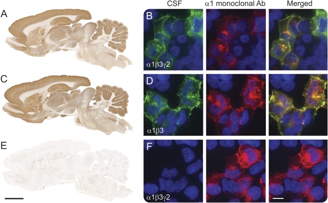
Methods: Clinical study of 26 patients, including 17 new (April 2013-January 2016) and 9 previously reported patients. Antibodies to α1, β3, and γ2 subunits of the GABAAR were determined using reported techniques.
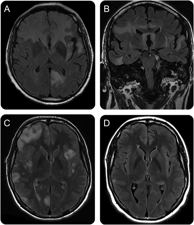
Results: Patients' median age was 40.5 years (interquartile range 48.5 [13.75-62.35] years; the youngest 2.5 months old; 13 female). Symptoms included seizures (88%), alteration of cognition (67%), behavior (46%), consciousness (42%), or abnormal movements (35%). Comorbidities were identified in 11 (42%) patients, including 7 tumors (mostly thymomas), 2 herpesvirus encephalitis (herpes simplex virus 1, human herpesvirus 6; coexisting with NMDAR antibodies), and 2 myasthenia without thymoma. Brain MRI was abnormal in 23 (88%) patients, showing in 20 (77%) multifocal, asynchronous, cortical-subcortical T2/fluid-attenuated inversion recovery abnormalities predominantly involving temporal (95%) and frontal (65%) lobes, but also basal ganglia and other regions. Immunologic or tumor therapy resulted in substantial improvement in 18/21 (86%) assessable patients; the other 3 (14%) died (2 status epilepticus, 1 sepsis). Compared with adults, children were more likely to have generalized seizures (p = 0.007) and movement disorders (p = 0.01) and less likely to have a tumor (p = 0.01). The main epitope targets were in the α1/β3 subunits of the GABAAR.
Conclusions: Anti-GABAAR encephalitis is characterized by frequent seizures and distinctive multifocal cortical-subcortical MRI abnormalities that provide an important clue to the diagnosis. The frequency of symptoms and comorbidities differ between children (more viral-related) and adults (more tumor-related). The disorder is severe but most patients respond to treatment.
© 2017 American Academy of Neurology.
Figures
Comment in
-
G2A1B3AA receptor antibodies and their clinical associations.Neurology. 2017 Mar 14;88(11):1010-1011. doi: 10.1212/WNL.0000000000003758. Epub 2017 Feb 15. Neurology. 2017. PMID: 28202698 No abstract available.
References
-
- Baulac S, Huberfeld G, Gourfinkel-An I, et al. First genetic evidence of GABA(A) receptor dysfunction in epilepsy: a mutation in the gamma2-subunit gene. Nat Genet 2001;28:46–48. - PubMed
-
- Wallace RH, Marini C, Petrou S, et al. Mutant GABAA receptor γ2-subunit in childhood absence epilepsy and febrile seizures. Nat Genet 2001;28:49–52. - PubMed
-
- Maljevic S, Krampfl K, Cobilanschi J, et al. A mutation in the GABA(A) receptor alpha(1)-subunit is associated with absence epilepsy. Ann Neurol 2006;59:983–987. - PubMed
-
- Lachance-Touchette P, Martin C, Poulin C, Gravel M, Carmant L, Cossette P. Screening of GABRB3 in French-Canadian families with idiopathic generalized epilepsy. Epilepsia 2010;51:1894–1897. - PubMed
MeSH terms
Substances
Grants and funding
LinkOut - more resources
Full Text Sources
Other Literature Sources
Medical
Miscellaneous