. 2017 Feb 28;114(9):2200-2205.
doi: 10.1073/pnas.1618307114. Epub 2017 Feb 15.

Stretchable living materials and devices with hydrogel-elastomer hybrids hosting programmed cells

Affiliations

Stretchable living materials and devices with hydrogel-elastomer hybrids hosting programmed cells

Xinyue Liu et al. Proc Natl Acad Sci U S A. .

Abstract

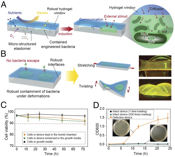
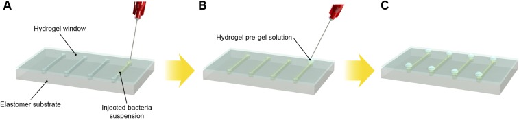
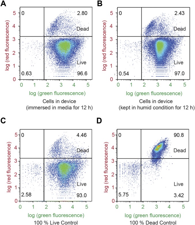
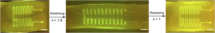
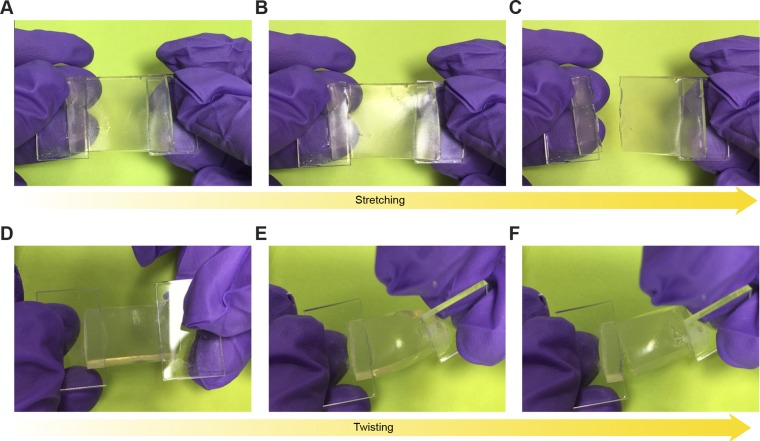
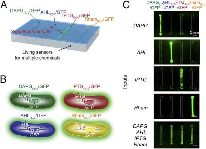
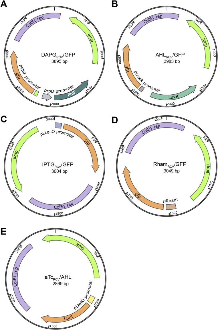
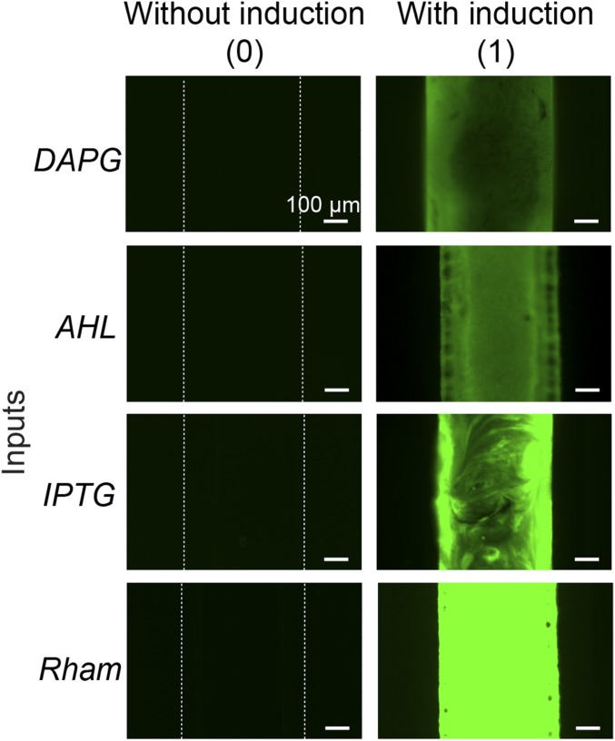
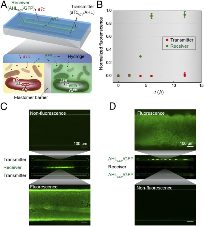
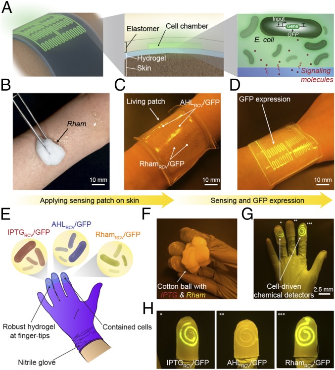
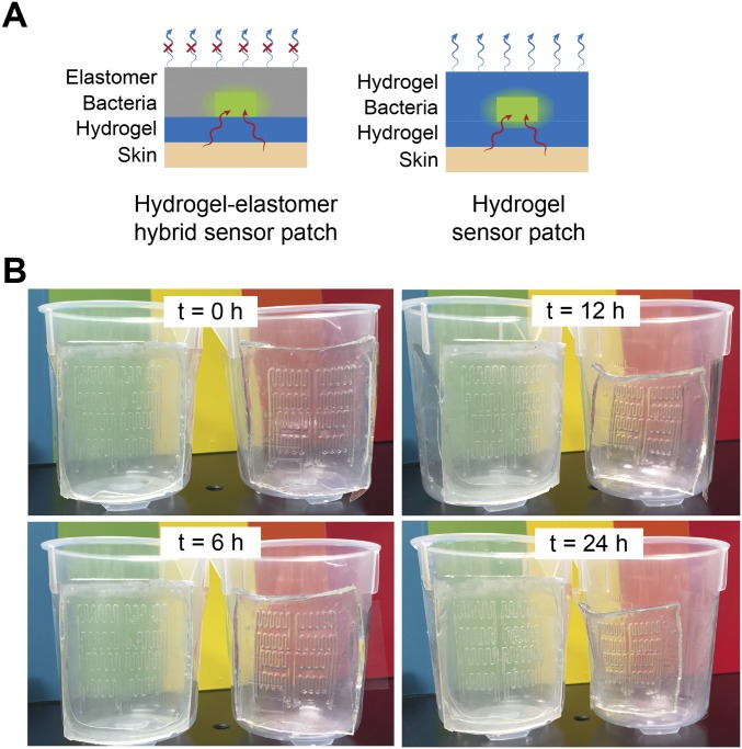
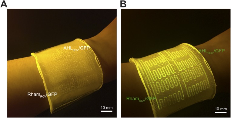
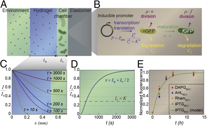
Living systems, such as bacteria, yeasts, and mammalian cells, can be genetically programmed with synthetic circuits that execute sensing, computing, memory, and response functions. Integrating these functional living components into materials and devices will provide powerful tools for scientific research and enable new technological applications. However, it has been a grand challenge to maintain the viability, functionality, and safety of living components in freestanding materials and devices, which frequently undergo deformations during applications. Here, we report the design of a set of living materials and devices based on stretchable, robust, and biocompatible hydrogel-elastomer hybrids that host various types of genetically engineered bacterial cells. The hydrogel provides sustainable supplies of water and nutrients, and the elastomer is air-permeable, maintaining long-term viability and functionality of the encapsulated cells. Communication between different bacterial strains and with the environment is achieved via diffusion of molecules in the hydrogel. The high stretchability and robustness of the hydrogel-elastomer hybrids prevent leakage of cells from the living materials and devices, even under large deformations. We show functions and applications of stretchable living sensors that are responsive to multiple chemicals in a variety of form factors, including skin patches and gloves-based sensors. We further develop a quantitative model that couples transportation of signaling molecules and cellular response to aid the design of future living materials and devices.

Keywords: biochemical sensors; genetically engineered bacteria; hydrogels; synthetic biology; wearable devices.


Conflict of interest statement

The authors declare no conflict of interest.

Figures

Fig. 1.
Fig. 1.
Design of living materials and devices. (A) Schematic illustration of a generic structure for living materials and devices. Layers of robust and biocompatible hydrogel and elastomer were assembled and bonded into a hybrid structure, which can transport sustained supplies of water, nutrient, and oxygen to genetically engineered cells at the hydrogel–elastomer interface. Communication between different types of cells and with the environment was achieved by diffusion of small molecules in hydrogels. (B) Schematic illustration of the high stretchability and high robustness of the hydrogel–elastomer hybrids that prevent cell leakage from the living device, even under large deformations. Images show that the living device can sustain uniaxial stretching over 1.8 times and twisting over 180° while maintaining its structural integrity. (C) Viability of bacterial cells at room temperature over 3 d. The cells were kept in the device placed in the humid chamber without additional growth media (yellow), in the device immersed in the growth media as a control (green), and in growth media as another control (black; n = 3 repeats). (D) OD600 and (Insets) streak plate results of the media surrounding the defective devices (yellow) and intact devices at different times after 1 (black) or 500 times (green) deformation of the living devices and immersion in media (n = 3 repeats).
Fig. S1.
Fig. S1.
Schematic illustration of cell suspension injection and sealing of injection points. (A) Bacteria were injected into the cavities at the hydrogel–elastomer interface with metallic needles from the hydrogel side. (B) Injection holes were sealed on the hydrogel–elastomer device with drops of fast-curable pregel solution. (C) We obtained the hydrogel–elastomer device with fully encapsulate bacteria.
Fig. S2.
Fig. S2.
Flow cytometry analysis using live/dead stains for (A) cells retrieved from the living device that has been immersed in media for 12 h, (B) cells retrieved from the living device that has been placed in humid environment for 12 h, (C) live-cell controls, and (D) dead-cell controls. Green fluorescence denotes both live and dead bacteria, and red fluorescence denotes bacteria that have been damaged and leaky membranes. The distributions of the live and dead populations are illustrated in the plots, with thresholds determined by controls. Over 95% of cells in the hydrogel–elastomer devices immersed in media or placed in humid chamber remained viable after 12 h.
Fig. S3.
Fig. S3.
Functional living device under large uniaxial stretch. After GFP was switched on in the wavy channels of Ecoflex–hydrogel hybrid matrix, the device was stretched to 1.8 times its original length and then released. The device, including cells encapsulated, can maintain functionality under large deformation without failure or leakage. (Scale bar: 5 mm.)
Fig. S4.
Fig. S4.
Deformation of agar-based living devices. An agar-based control device that encapsulated RhamRCV/GFP bacteria with the same dimensions as the hydrogel–elastomer hybrid was fabricated. The agar device fractured even under moderate deformation, including (A–C) a stretch of 1.1 or (DF) a twist of 60°.
Fig. S5.
Fig. S5.
Cell leakage from the agar device. The medium surrounding the agar device (without any deformation) was collected to measure OD600. The high OD600 after 10 h indicates the large cell populations in the medium and cell leakage even without any deformation of agar gel.
Fig. 2.
Fig. 2.
Stretchable living sensors can independently detect multiple chemicals. (A) Schematic illustration of a hydrogel–elastomer hybrid with four isolated chambers to host bacterial strains, including DAPGRCV/GFP, AHLRCV/GFP, IPTGRCV/GFP, and RhamRCV/GFP. Signaling molecules were diffused from the environment through the hydrogel window into cell chambers, where they were detected by the bacteria. (B) Genetic circuits were constructed in bacterial strains to detect cognate inducers (i.e., DAPG, AHL, IPTG, and Rham) and produce GFP. (C) Images of living devices after exposure to individual or multiple inputs. Cell chambers hosting bacteria with the cognate sensors showed green fluorescence, whereas the noncognate bacteria in chambers were not fluorescent. Scale bars are shown in images.
Fig. S6.
Fig. S6.
Plasmid maps of the plasmids constructed. (A) DAPGRCV/GFP, (B) AHLRCV/GFP, (C) IPTGRCV/GFP, (D) RhamRCV/GFP, and (E) aTcRCV/AHL. Plasmids were constructed as described in SI Text. amp, Ampicillin resistance gene; ColE1 rep:, replication origin from ColE1 plasmid.
Fig. S7.
Fig. S7.
Microscopic images of different cell strains in the chamber encapsulated in the living device. When a cell strain was induced, the channels showed fluorescence [denoted as (1)]. If not induced, the channel stayed dark [denoted as (0)]. Scale bars are shown in images.
Fig. 3.
Fig. 3.
Interactive genetic circuits. (A) Schematic illustration of a living device that contains two cell strains: the transmitters (aTcRCV/AHL strain) produce AHL in the presence of aTc, and the receivers (AHLRCV/GFP strain) express GFP in the presence of AHL. The transmitters could communicate with the receivers via diffusion of the AHL signaling molecules through the hydrogel window, although the cells are physically isolated by elastomer. (B) Quantification of normalized fluorescence over time (n = 3 repeats). All data were measured by flow cytometry, with cells retrieved from the device at different times. (C) Images of device and microscopic images of cell chambers 6 h after addition of aTc into the environment surrounding the device. The side chambers contain transmitters, whereas the middle one contains receivers. (D) Images of device and microscopic images of cell chambers 6 h after aTc addition in the environment. The side chambers contain aTcRCV/GFP instead of transmitters, whereas the middle one contains receivers. Scale bars are shown in images.
Fig. 4.
Fig. 4.
Living wearable devices. (A) Schematic illustration of a living patch. The patch adhered to the skin with the hydrogel side, and the elastomer side was exposed to the air. Engineered bacteria inside can detect signaling molecules. (B–D) Rham solution was smeared on skin, and the sensor patch was conformably applied on skin. The channels with RhamRCV/GFP in the living patch became fluorescent, whereas channels with AHLRCV/GFP did not show any differences. Scale bars are shown in images. (E) Schematic illustration of a glove with chemical detectors robustly integrated at the fingertips. Different chemical-inducible cell strains, including IPTGRCV/GFP, AHLRCV/GFP, and RhamRCV/GFP, were encapsulated in the chambers. (F–H) When the living glove was used to grab a wet cotton ball containing the inducers, GFP fluorescence was shown in the cognate sensors IPTGRCV/GFP (*) and RhamRCV/GFP (***) on the gloves. In contrast, the noncognate sensor AHLRCV/GFP (**) did not show any fluorescence. Scale bars are shown in images.
Fig. S8.
Fig. S8.
Antidehydration property of the sensor patch. (A) Schematic illustration of the hydrogel–elastomer hybrid sensor patch, which has the antidehydration property over the pure hydrogel device. The silicone elastomer cover effectively prevents evaporation of water from the hydrogel and dehydration of the living patch. (B) Time-lapse snapshots of hydrogel–elastomer hybrid sensor patch (Left) and pure hydrogel sensor patch (Right) mounted on a plastic beaker at room temperature with low humidity (25 °C and 50% relative humidity) for 24 h. The elastomer outer layer of the hydrogel–elastomer hybrid device significantly slowed down the dehydration process of the hydrogel and provided a sustained humid environment for encapsulated cells for over 24 h. However, distorted channels became apparent on patches made of pure hydrogels when they were exposed to air for 6 h because of dehydration.
Fig. S9.
Fig. S9.
Living patch control experiments. (A) When no inducer was smeared on skin and the living sensor patch was adhered on skin conformably, the channels with RhamRCV/GFP and AHLRCV/GFP in the living patch did not show any differences. (B) When both inducers Rham and AHL were smeared on skin and the living patch was applied, the channels with RhamRCV/GFP and AHLRCV/GFP in the living patch became fluorescent. Scale bars are shown in images.
Fig. 5.
Fig. 5.
Model for the diffusion–induction process in living materials and devices. (A) Schematic illustration of the diffusion of signaling molecules from the environment through the hydrogel to cell chambers in the living device. (B) Diagram of GFP expression after induction with a small molecule chemical. (C) Inducer concentration profile throughout the hydrogel window and cell chamber at different times. (D) Typical inducer concentration in the cell chamber as a function of time. (E) The normalized fluorescence of different cell strains as a function of time after addition of inducer (n = 3 repeats). Dots represent experimental data, and curve represents the model.
Fig. S10.
Fig. S10.
Calculation of critical diffusion timescales for living materials and devices. (A) Schematic illustration of signaling molecule diffusion from the environment through the hydrogel in the living device. Cells were embedded in a segment of the hydrogel close to the elastomer wall. (B) Comparison of typical inducer concentration profiles when cells were embedded in hydrogel [I(L,t)/I0] vs. cells in medium of the cell chamber [I(Lg+Lc/2,t)/I0]. Despite small deviation (<12%) because of the diffusivity differences between hydrogel and medium and distance variation in two cases, it can be seen that the profile in the simplified model can consistently represent the typical concentration profile in the cell chamber of the living sensor at any time.
Fig. S11.
Fig. S11.
Approximate diffusion timescale. The expression of K/I0=2erfc[L/2Dgt]erfc[L/Dgt] and its approximate solution. The prefactor in tdiffuse=[Λ(K/I0)]2L2/Dg is fitted into a power law that approximately gives tdiffuse4/9(I0/K1)0.56L2/Dg.

References

    1. Pardee K, et al. Paper-based synthetic gene networks. Cell. 2014;159(4):940–954. - PMC - PubMed
    1. Mimee M, Tucker AC, Voigt CA, Lu TK. Programming a human commensal bacterium, Bacteroides thetaiotaomicron, to sense and respond to stimuli in the murine gut microbiota. Cell Syst. 2015;1(1):62–71. - PMC - PubMed
    1. Friedland AE, et al. Synthetic gene networks that count. Science. 2009;324(5931):1199–1202. - PMC - PubMed
    1. Siuti P, Yazbek J, Lu TK. Engineering genetic circuits that compute and remember. Nat Protoc. 2014;9(6):1292–1300. - PubMed
    1. Chen AY, et al. Synthesis and patterning of tunable multiscale materials with engineered cells. Nat Mater. 2014;13(5):515–523. - PMC - PubMed

Publication types

MeSH terms