. 2017 Feb 28;114(9):2247-2252.
doi: 10.1073/pnas.1609243114. Epub 2017 Feb 15.

Atomic structure of granulin determined from native nanocrystalline granulovirus using an X-ray free-electron laser

Affiliations

Atomic structure of granulin determined from native nanocrystalline granulovirus using an X-ray free-electron laser

Cornelius Gati et al. Proc Natl Acad Sci U S A. .

Abstract

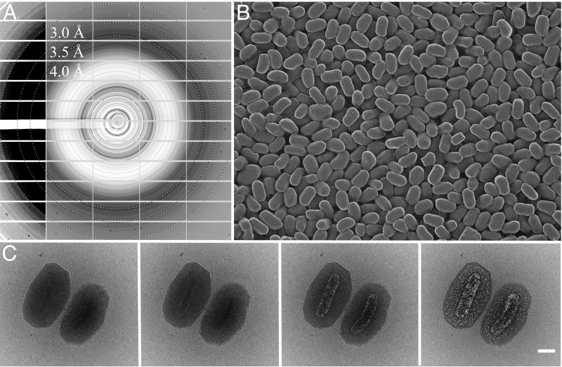
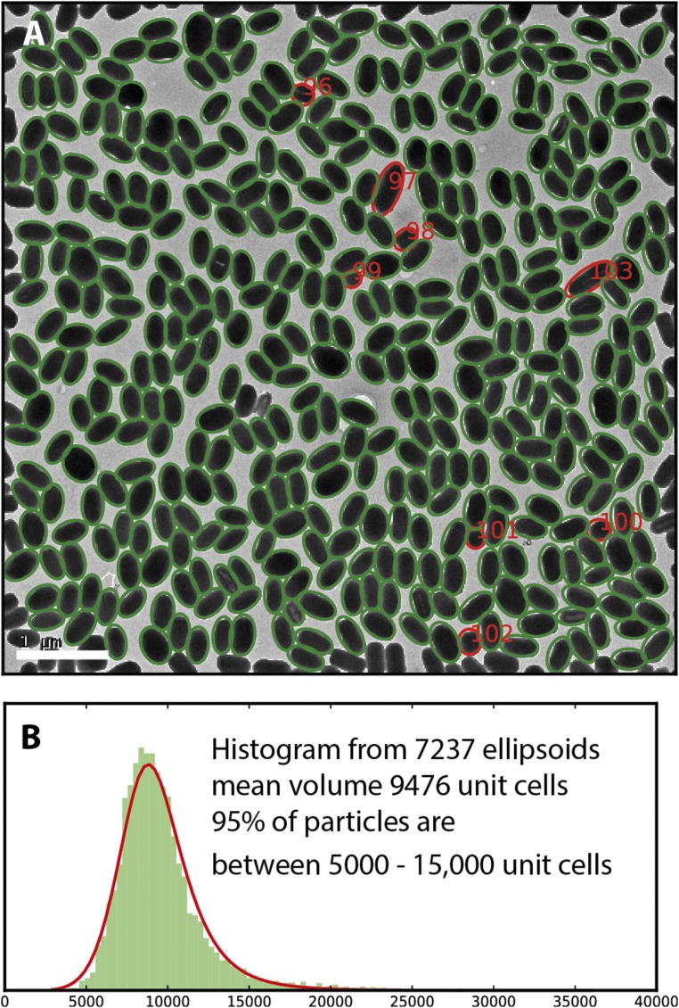
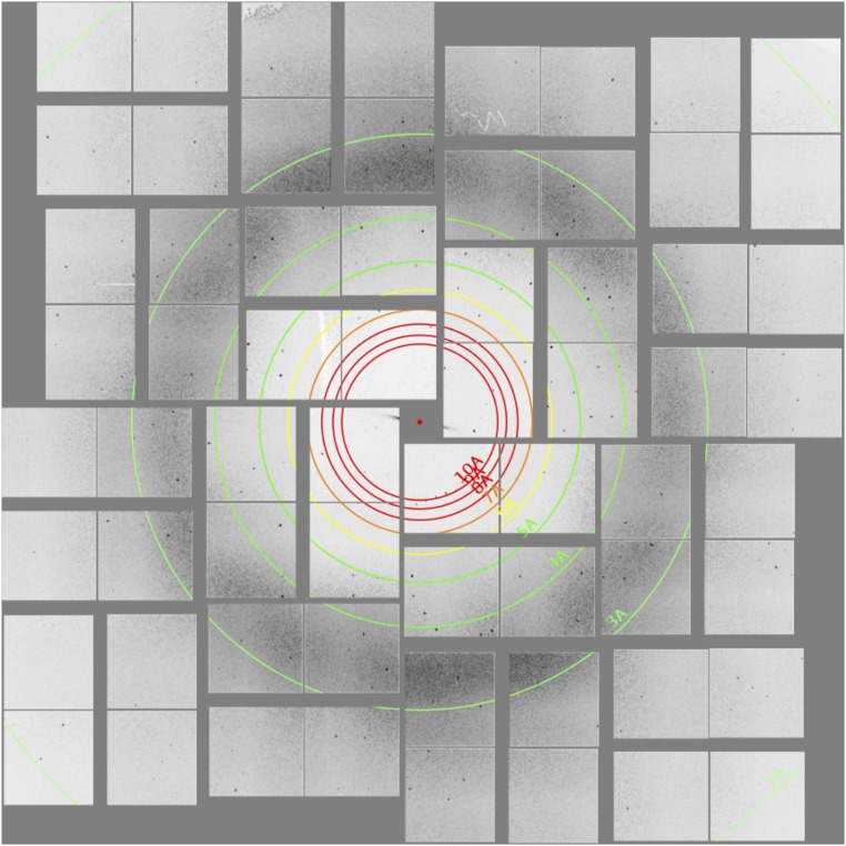
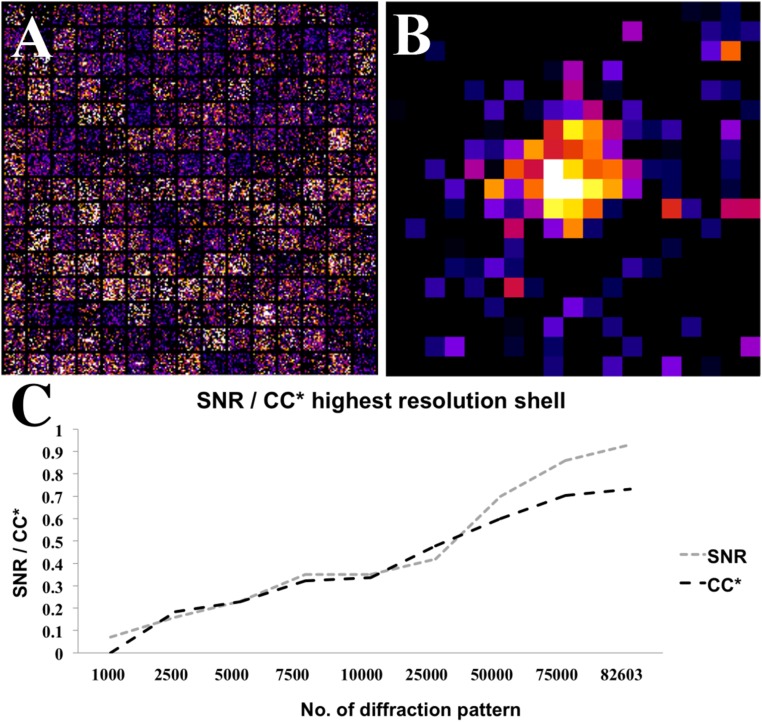
To understand how molecules function in biological systems, new methods are required to obtain atomic resolution structures from biological material under physiological conditions. Intense femtosecond-duration pulses from X-ray free-electron lasers (XFELs) can outrun most damage processes, vastly increasing the tolerable dose before the specimen is destroyed. This in turn allows structure determination from crystals much smaller and more radiation sensitive than previously considered possible, allowing data collection from room temperature structures and avoiding structural changes due to cooling. Regardless, high-resolution structures obtained from XFEL data mostly use crystals far larger than 1 μm3 in volume, whereas the X-ray beam is often attenuated to protect the detector from damage caused by intense Bragg spots. Here, we describe the 2 Å resolution structure of native nanocrystalline granulovirus occlusion bodies (OBs) that are less than 0.016 μm3 in volume using the full power of the Linac Coherent Light Source (LCLS) and a dose up to 1.3 GGy per crystal. The crystalline shell of granulovirus OBs consists, on average, of about 9,000 unit cells, representing the smallest protein crystals to yield a high-resolution structure by X-ray crystallography to date. The XFEL structure shows little to no evidence of radiation damage and is more complete than a model determined using synchrotron data from recombinantly produced, much larger, cryocooled granulovirus granulin microcrystals. Our measurements suggest that it should be possible, under ideal experimental conditions, to obtain data from protein crystals with only 100 unit cells in volume using currently available XFELs and suggest that single-molecule imaging of individual biomolecules could almost be within reach.

Keywords: SFX; XFEL; bioimaging; nanocrystals; structural biology.


Conflict of interest statement

The authors declare no conflict of interest.

Figures

Fig. 1.
Fig. 1.
Granulovirus OBs contain a single virion surrounded by a crystalline protein layer that diffracts to high resolution. (A) Powder X-ray diffraction from a pellet of granulovirus OBs at 100 K (Materials and Methods). Protein diffraction rings extend to a resolution between 3 and 3.5 Å. The detector panels on the left with enhanced contrast show evidence of diffraction at even higher resolution. Resolution rings are shown at 4, 3.5, and 3 Å. (B) Freeze etch electron micrograph showing the uniform size distribution of the particles (Materials and Methods). (C) Cryo-EM. The sequence of four 20 e/Å2 exposures shows the effects of radiation damage on granulovirus OBs. The crystalline lattice is visible only in the first image and hydrogen gas bubbles produced by radiolysis eventually reveal the virion. (Scale bar, 100 nm.)
Fig. S1.
Fig. S1.
(A) Electron micrograph showing granulovirus occlusion bodies outlined by ellipses. The particles shown with red ellipses are outliers. Particles 97 and 103 are unusally large and the other red particles unusually small, probably because they are end-on views. (B) Histogram of volumes derived from 7,237 ellipsoids from particles in 16 similar images. The histogram has been fitted with a log-logistic distribution with mode ∼9,000 unit cells. According to this distribution, 2% of the particles are larger than 16,000 unit cells. (Scale bar, 1 µm.)
Fig. S2.
Fig. S2.
Example of a high-resolution diffraction pattern collected on the CSPAD, showing intense diffraction into the corners of the detector.
Fig. 2.
Fig. 2.
Averaging thousands of reflections significantly improved overall resolution estimate. (A) Two hundred and twenty-five randomly selected single images at the predicted location of the 21 26 29 reflection (corresponding to 2.3 Å resolution). (B) Averaged Bragg intensity of the 21 26 29 reflection intensities from 3,176 observed reflections after first rotating them into the common frame of reference of the lattice. (C) Plot of overall signal-to-noise ratio and CC* metric in the 2–2.1 Å resolution shell against the number of indexed diffraction patterns included.
Fig. S3.
Fig. S3.
Results of the “detwinning” step of the CpGV dataset by CrystFEL, showing intensities in the hk0 plane after merging symmetry-equivalent reflections in point group m3¯ (A) before applying ambigator showing an additional, artificial symmetry axis resulting in a fourfold symmetry of the hkl intensities and (B) after ambigator showing the true twofold symmetry.
Fig. S4.
Fig. S4.
Quality metrics of the final dataset. (A) Correlation coefficient of all indexed patterns to an initially merged set against individual scaling factors. (B) Rsplit vs. number of patterns. (C) CC* vs. number of patterns. (D) Number of patterns vs. average Bragg peak intensity.
Fig. 3.
Fig. 3.
(A) The structure of the biological unit of granulin building blocks forming the crystalline OB. (B) Granulin monomer. The SFX structure is displayed in blue and the SYN structure in green. Regions of granulin that were present in the SFX but not in the SYN structure are highlighted in red.
Fig. 4.
Fig. 4.
Comparison of the final 2Fo-Fc electron density map and model for the SFX (blue) and SYN (green) structures of granulin. (A) SFX and (B) SYN model with color-map and thickness corresponding to local B-factor. (C and D) Close-up of Cys135 in (C) the SFX (blue) and (D) the SYN structure and (green) embedded in its electron (gray mesh, contoured 1.5 σ). Cys135 adopts two distinct side chain conformations (shown as sticks): The major conformer forms an intermolecular disulfide bond with its counterpart in a neighboring molecule and has a refined occupancy of 0.68 and 0.87 for the SFX and SYN structure, respectively. (E) SFX and (F) SYN comparison of electron density for residues 168–201 (contoured at 1 σ). Electron density defining residues 176–190 was only found for the SFX structure.
Fig. S5.
Fig. S5.
Simulated diffraction patterns of granulin nanocrystals with 9,000 unit cells (A) and 123 unit cells (B). A dose of 1 GGy—color bar indicates the photon count in each pixel. The Inset illustrates the model of the crystals used in the simulations.

References

    1. Henderson R. The potential and limitations of neutrons, electrons and X-rays for atomic resolution microscopy of unstained biological molecules. Q Rev Biophys. 1995;28(2):171–193. - PubMed
    1. Howells MR, et al. An assessment of the resolution limitation due to radiation-damage in x-ray diffraction microscopy. J Electron Spectrosc Relat Phenom. 2009;170(1-3):4–12. - PMC - PubMed
    1. Holton JM. A beginner’s guide to radiation damage. J Synchrotron Radiat. 2009;16(Pt 2):133–142. - PMC - PubMed
    1. Warkentin M, Hopkins JB, Haber JB, Blaha G, Thorne RE. Temperature-dependent radiation sensitivity and order of 70S ribosome crystals. Acta Crystallogr D Biol Crystallogr. 2014;70(Pt 11):2890–2896. - PMC - PubMed
    1. Henderson R. Cryo-protection of protein crystals against radiation damage in electron and X-ray diffraction. Proc Biol Sci. 1990;241(1300):6–8.

Publication types

Substances

LinkOut - more resources