. 2017 Feb 16:7:42788.
doi: 10.1038/srep42788.

Calpain-1 deletion impairs mGluR-dependent LTD and fear memory extinction

Affiliations

Calpain-1 deletion impairs mGluR-dependent LTD and fear memory extinction

Guoqi Zhu et al. Sci Rep. .

Abstract

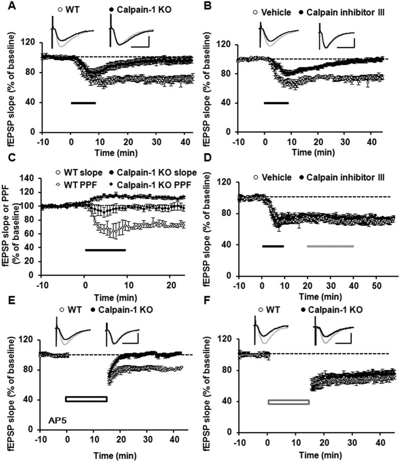
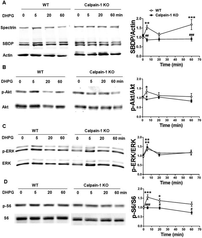
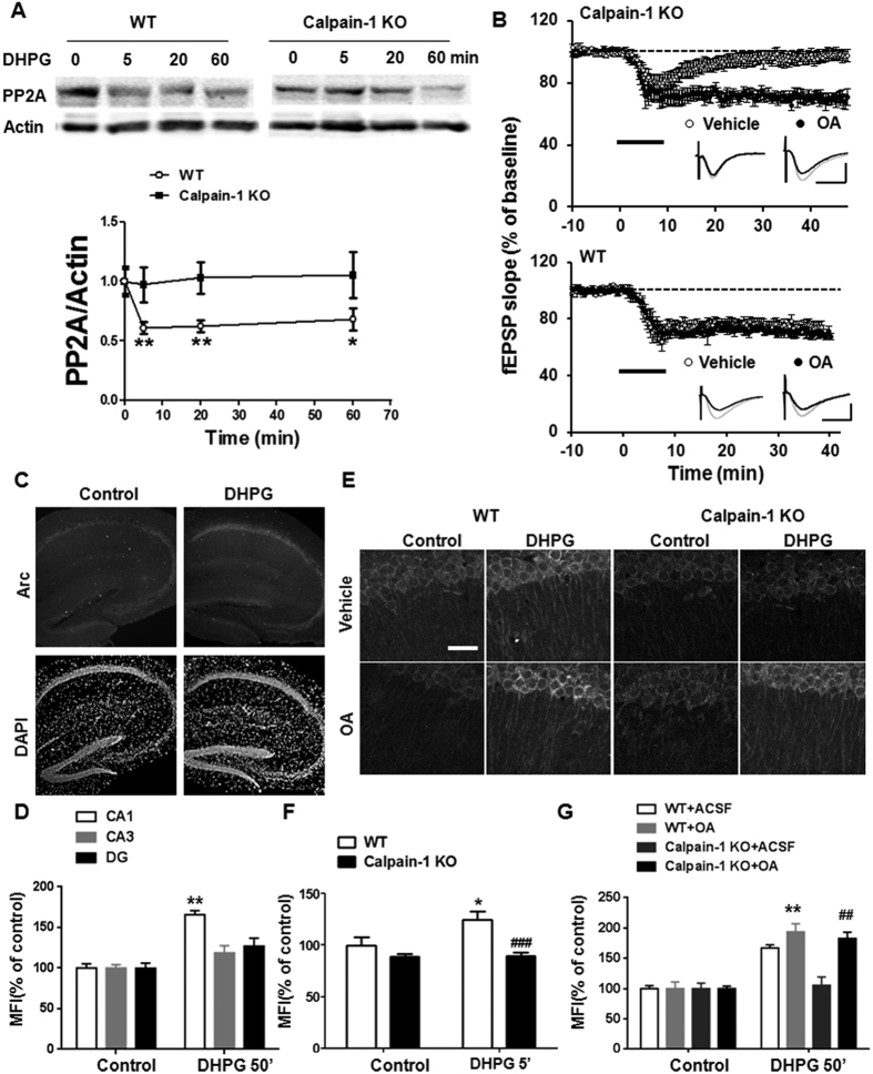
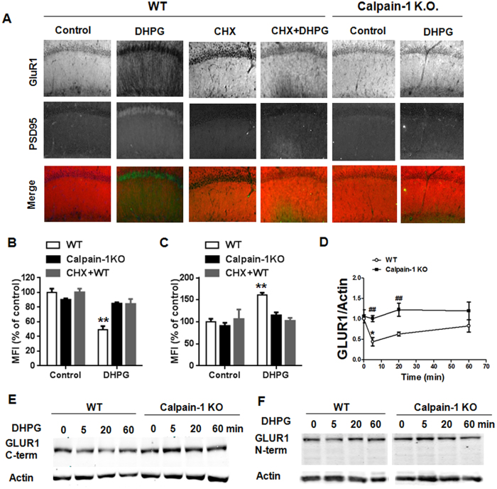
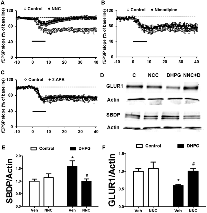
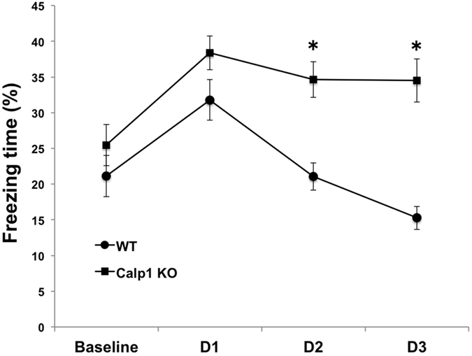
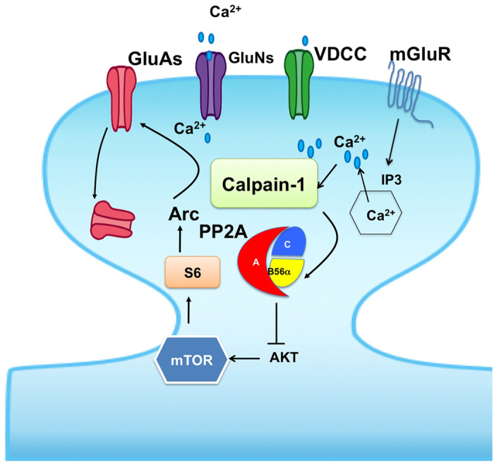
Recent studies indicate that calpain-1 is required for the induction of long-term potentiation (LTP) elicited by theta-burst stimulation in field CA1 of hippocampus. Here we determined the contribution of calpain-1 in another type of synaptic plasticity, the long-term depression (LTD) elicited by activation of type-I metabotropic glutamate receptors (mGluR-LTD). mGluR-LTD was associated with calpain-1 activation following T-type calcium channel opening, and resulted in the truncation of a regulatory subunit of PP2A, B56α. This signaling pathway was required for both the early and late phase of Arc translation during mGluR-LTD, through a mechanism involving mTOR and ribosomal protein S6 activation. In contrast, in hippocampal slices from calpain-1 knock-out (KO) mice, application of the mGluR agonist, DHPG, did not result in B56α truncation, increased Arc synthesis and reduced levels of membrane GluA1-containing AMPA receptors. Consistently, mGluR-LTD was impaired in calpain-1 KO mice, and the impairment could be rescued by phosphatase inhibitors, which also restored Arc translation in response to DHPG. Furthermore, calpain-1 KO mice exhibited impairment in fear memory extinction to tone presentation. These results indicate that calpain-1 plays a critical role in mGluR-LTD and is involved in many forms of synaptic plasticity and learning and memory.


Conflict of interest statement

The authors declare no competing financial interests.

Figures

Figure 1
Figure 1. Calpain-1 deletion impairs DHPG-induced LTD.
(A) DHPG application (100 μM for 10 min, horizontal bar) resulted in LTD in field CA1 of WT mice but the slopes of field excitatory postsynaptic potential (fEPSP) returned to baseline within 10 min in slices from calpain-1 KO mice. (B) Application of calpain inhibitor III (10 μM) during DHPG application inhibited DHPG-induced LTD in hippocampal slices from WT mice. (C) DHPG-induced enhancement of paired pulse facilitation (PPF) was not affected in hippocampal slices from calpain-1 KO mice. (D) Application of calpain inhibitor III (10 μM) 10 min after DHPG treatment did not modify mGluR-LTD. (E) LTD elicited by PP-LFS in the presence of AP5 (50 μM) was also impaired in calpain-1 KO mice. (F) On the other hand, LFS-induced LTD was not affected in hippocampal slices from calpain-1 KO mice, and was identical to that in slices from WT mice. Results are means ± S.E.M. of 5–6 slices from 4 different animals. Grey and black traces represent responses during baseline and 40 min after DHPG application, respectively. Scale bar: 0.5 mV/10 ms.
Figure 2
Figure 2. DHPG application triggers calpain-1 activation and Akt and S6 phosphorylation.
Hippocampal slices were treated with DHPG (100 μM, for 10 min). Slices were collected at various times after DHPG application, homogenized, and aliquots of the homogenates were processed for western blots labeled with the indicated antibodies, as described in Materials and Methods. (A–D) Left panel: Representative blots of spectrin and spectrin breakdown products (SBDP) (A), phospho-Akt (pAkt) and Akt (B), p-ERK and ERK (C), p-S6 and S6 (D) after DHPG application in hippocampal slices from WT and calpain-1 KO mice. Right panel: Quantification of the ratios of the indicated proteins after DHPG application in WT and calpain-1 KO mice. In all cases, results are means ± S.E.M. of 4–7 slices from 4 different animals; *p < 0.05, **p < 0.01, ***p < 0.001, as compared with baseline values (time 0), #p < 0.05, ##p < 0.01, ###p < 0.001, as compared with the corresponding time point in WT mice (Two-way ANOVA + Bonferroni test).
Figure 3
Figure 3. DHPG application results in calpain-1-mediated B56α degradation and PP2A inhibition restores DHPG-induced Arc translation and LTD in calpain-1 KO mice.
(A) DHPG application in hippocampal slices from WT mice but not from calpain-1 KO mice elicited a rapid decrease in B56α levels. Upper panel: representative blots for B56α after DHPG application in both of WT and calpain-1 KO mice. Bottom panel: quantification of B56α/actin ratio. Results are means ± S.E.M. of 6–9 slices from 4 different animals; *p < 0.05, **p < 0.01, as compared with baseline value (time 0) in WT mice; #p < 0.05, as compared with corresponding time point in WT mice (Two-way ANOVA + Bonferroni test). (B) The PP2A inhibitor okadaic acid (OA, 50 nM) restored mGluR-LTD in hippocampal slices from calpain-1 KO mice (top), but did not affect mGluR-LTD in slices from WT mice (bottom). Grey and black traces represent responses during baseline and 40 min after DHPG application, respectively. Scale bar: 0.5 mV/10 ms. Results are means ± S.E.M. of 5–8 slices from 5 different animals. (C) In WT mice, DHPG application (100 μM for 10 min) induced a significant increase in Arc levels in CA1 region of hippocampus but not in CA3 or dentate gyrus (DG) at 50 min after DHPG application. Upper panel shows representative images of Arc immunostaining. Bottom panel shows the images of DAPI staining. (D) Quantification data of the mean fluorescence intensity (MFI) in different regions of hippocampus; **p < 0.01, as compared with baseline level in WT mice (Two-way ANOVA + Bonferroni test). (E) DHPG induced a late (50 min after DHPG) increase in Arc immunostaining in hippocampal slices from WT mice, which was restored by okadaic acid (OA) application in calpain-1 KO mice. (F,G) Quantification data of the MFI of Arc immunostaining (somatic and dendritic) after treatment with DHPG for 5 (F) or 50 min (G). (F) *p < 0.05 compared with control in WT mice. ###p < 0.001 compared with DHPG in WT mice (Two-way ANOVA + Bonferroni test). (G) **p < 0.01 compared with DHPG in WT mice. ##p < 0.01 compared with DHPG in calpain-1 KO mice (Two-way ANOVA + Bonferroni test). In all cases, results are means ± S.E.M. of 5–10 slices from 5 different animals.
Figure 4
Figure 4. DHPG application induces a protein synthesis-dependent decrease in GluA1 and increase in PSD95.
(A) Representative images showing that DHPG application (100 μM, for 10 min) causes a remarkable decrease in GluA1 expression and increase of PSD95 in CA1 region of WT mice 50 min after DHPG application, which were blocked by cycloheximide (CHX, 25 μM). In calpain-1 KO mice, DHPG did not affect GluA1 or PSD95 immunostaining. (B,C) Quantification data of mean fluorescence intensity (MFI) of GluA1 (B) and PSD95 (C) immunostaining; **p < 0.01, as compared with the corresponding control (Two-way ANOVA + Bonferroni test). (D) Quantification data of GluA1/actin ratio; *p < 0.05, as compared with baseline level; ##p < 0.01, as compared with the corresponding time point in WT mice (Two-way ANOVA + Bonferroni test). (E,F) Representative blots of GluA1 at various time points after DHPG application in WT and calpain-1 KO mice using a C-terminal (E) or an N-terminal (F) GluA1 antibody. In all cases, results are means ± S.E.M. of 4–8 slices from 4 different animals.
Figure 5
Figure 5. DHPG-induced GluA1 down-regulation and LTD is suppressed by a T-type voltage dependent calcium channel blocker.
(A) The T-type voltage dependent calcium channel inhibitor NCC (10 μM) blocked DHPG-induced LTD. (B) The L-type voltage dependent calcium channel inhibitor nimodipine (10 μM) did not affect LTD. (C) The IP3 channel inhibitor 2-APB (50 μM) did not affect DHPG-induced LTD. (D) NCC inhibited DHPG-induced increase in SBDP and decrease in GLUA1. (E,F) Quantification data of SBDP/actin (E) or GluA1/actin (F) ratio after 5 min of DHPG treatment in hippocampal slices from WT mice; *p < 0.05, as compared with vehicle control; ##p < 0.01, as compared with vehicle DHPG (Two-way ANOVA + Bonferroni test). In all cases, results are means ± S.E.M. of 5–8 slices from 5 different animals.
Figure 6
Figure 6. Calpain-1 KO mice exhibit impaired extinction of fear memory.
Groups of WT and calpain-1 KO mice were trained in the fear conditioning protocol with 3 tone-shock pairings 1 min apart. Twenty-four h later, they were placed in a different context and exposed to repeated exposure to tone without shock (14 trials with 5 sec tone exposure with an inter-trial interval of 25 sec). This protocol was repeated for 2 more days for a total of 3 days of extinction. The percent of freezing following each tone presentation was determined and the cumulative percent freezing time per day determined and averaged. Results are means ± S.E.M. of 15–20 animals per group. **p < 0.01, as compared with the corresponding time point in WT mice (Two-way ANOVA + Bonferroni test).
Figure 7
Figure 7. Schematic illustration of the potential mechanism underlying mGluR- LTD.
In the proposed model, mGluR1/5 stimulation activates calpain-1 following calcium entry through T-type voltage dependent calcium channel. Calpain-1 degrades the B56α subunit of PP2A, which results in decreased PP2A activity and stimulation of the Akt/mTOR/S6 pathway, leading to increase in Arc and PSD95 synthesis. Increased synaptic levels of Arc and PSD95 facilitate endocytosis of GluA1-containing AMPA receptors, producing LTD. In calpain-1 KO mice, the lack of B56α degradation in response to mGluR1/5 stimulation prevents activation of this signaling pathway, which results in LTD impairment. Application of a PP2A inhibitor substitutes for calpain-1 activation, and therefore reverses the impairment in LTD and the associated changes in Arc and PSD95.

Similar articles

Cited by

References

    1. Neves G., Cooke S. F. & Bliss T. V. Synaptic plasticity, memory and the hippocampus: a neural network approach to causality. Nat. Rev. Neurosci. 9, 65–75 (2008). - PubMed
    1. Kennedy M. B. Synaptic Signaling in Learning and Memory. Cold Spring Harbor Perspect. Biol. 8, a016824 (2016). - PMC - PubMed
    1. Sweatt J. D. Neural Plasticity & Behavior - Sixty Years of Conceptual Advances. J. Neurochem. Epub ahead of print (2016). - PubMed
    1. Kim J. et al.. Blockade of amygdala metabotropic glutamate receptor subtype 1 impairs fear extinction. Biochem. Biophys. Res. Comm. 355, 188–193 (2007). - PubMed
    1. Dalton G. L., Wang Y. T., Floresco S. B. & Phillips A. G. Disruption of AMPA receptor endocytosis impairs the extinction, but not acquisition of learned fear. Neuropsychopharmacology 33, 2416–2426 (2008). - PubMed

Publication types