

Quality Assurance using Outlier Detection on an Automatic Segmentation Method for the Cerebellar Peduncles
- PMID: 28203039
- PMCID: PMC5304449
- DOI: 10.1117/12.2217309
Quality Assurance using Outlier Detection on an Automatic Segmentation Method for the Cerebellar Peduncles
Abstract
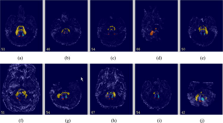
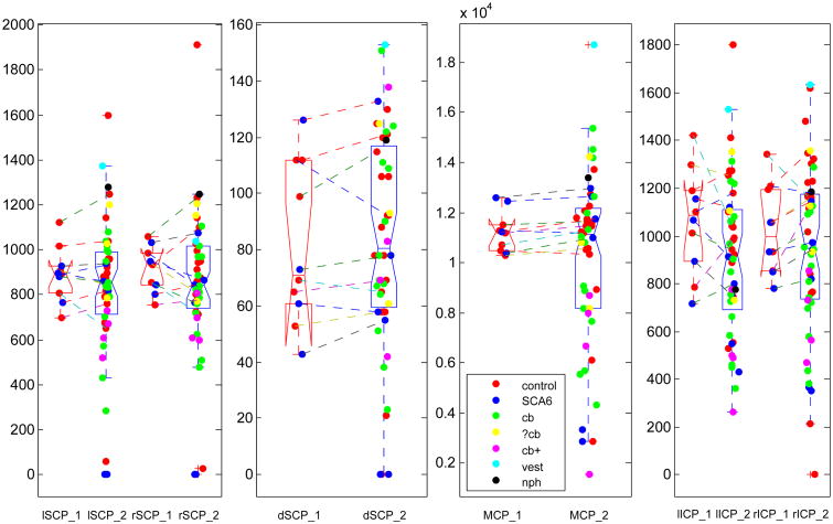
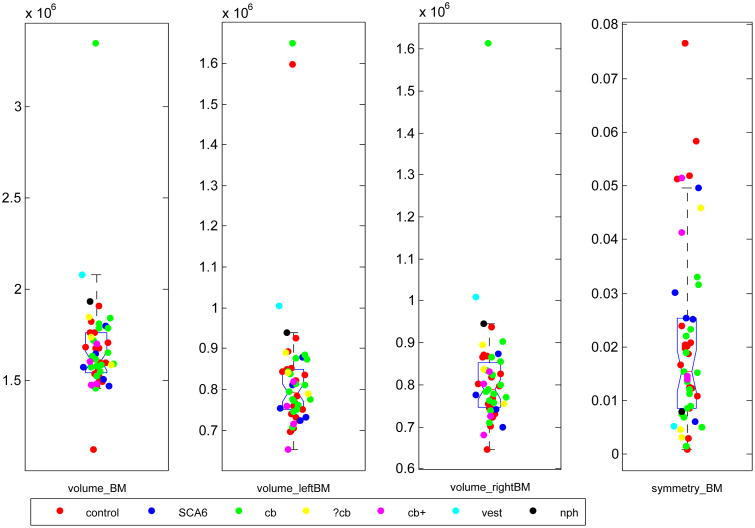
Cerebellar peduncles (CPs) are white matter tracts connecting the cerebellum to other brain regions. Automatic segmentation methods of the CPs have been proposed for studying their structure and function. Usually the performance of these methods is evaluated by comparing segmentation results with manual delineations (ground truth). However, when a segmentation method is run on new data (for which no ground truth exists) it is highly desirable to efficiently detect and assess algorithm failures so that these cases can be excluded from scientific analysis. In this work, two outlier detection methods aimed to assess the performance of an automatic CP segmentation algorithm are presented. The first one is a univariate non-parametric method using a box-whisker plot. We first categorize automatic segmentation results of a dataset of diffusion tensor imaging (DTI) scans from 48 subjects as either a success or a failure. We then design three groups of features from the image data of nine categorized failures for failure detection. Results show that most of these features can efficiently detect the true failures. The second method-supervised classification-was employed on a larger DTI dataset of 249 manually categorized subjects. Four classifiers-linear discriminant analysis (LDA), logistic regression (LR), support vector machine (SVM), and random forest classification (RFC)-were trained using the designed features and evaluated using a leave-one-out cross validation. Results show that the LR performs worst among the four classifiers and the other three perform comparably, which demonstrates the feasibility of automatically detecting segmentation failures using classification methods.
Keywords: box-whisker plot; cerebellar peduncles; classification; outlier detection; quality assurance; segmentation.
Figures


Similar articles
-
Segmentation of the Cerebellar Peduncles Using a Random Forest Classifier and a Multi-object Geometric Deformable Model: Application to Spinocerebellar Ataxia Type 6.Neuroinformatics. 2015 Jul;13(3):367-81. doi: 10.1007/s12021-015-9264-7. Neuroinformatics. 2015. PMID: 25749985 Free PMC article.
-
Automatic white matter lesion segmentation using an adaptive outlier detection method.Magn Reson Imaging. 2012 Jul;30(6):807-23. doi: 10.1016/j.mri.2012.01.007. Epub 2012 May 11. Magn Reson Imaging. 2012. PMID: 22578927
-
Lumen-intima and media-adventitia segmentation in IVUS images using supervised classifications of arterial layers and morphological structures.Comput Methods Programs Biomed. 2019 Aug;177:113-121. doi: 10.1016/j.cmpb.2019.05.021. Epub 2019 May 21. Comput Methods Programs Biomed. 2019. PMID: 31319939
-
A self-supervised strategy for fully automatic segmentation of renal dynamic contrast-enhanced magnetic resonance images.Med Phys. 2019 Oct;46(10):4417-4430. doi: 10.1002/mp.13715. Epub 2019 Aug 16. Med Phys. 2019. PMID: 31306492
-
Cavity contour segmentation in chest radiographs using supervised learning and dynamic programming.Med Phys. 2014 Jul;41(7):071912. doi: 10.1118/1.4881096. Med Phys. 2014. PMID: 24989390
Cited by
-
Liver Function Biomarkers and Lung Cancer Risk: A Prospective Cohort Study in the UK Biobank.Clin Respir J. 2024 Dec;18(12):e70042. doi: 10.1111/crj.70042. Clin Respir J. 2024. PMID: 39721983 Free PMC article.
-
Comparing fully automated state-of-the-art cerebellum parcellation from magnetic resonance images.Neuroimage. 2018 Dec;183:150-172. doi: 10.1016/j.neuroimage.2018.08.003. Epub 2018 Aug 9. Neuroimage. 2018. PMID: 30099076 Free PMC article.
References
-
- Sivaswamy Lalitha, et al. A diffusion tensor imaging study of the cerebellar pathways in children with autism spectrum disorder. Journal of child neurology. 2010 - PubMed
-
- Le Bihan Denis, et al. Diffusion tensor imaging: concepts and applications. Journal of magnetic resonance imaging. 2001;13.4:534–546. - PubMed
-
- Ye Chuyang, et al. Labeling of the cerebellar peduncles using a supervised Gaussian classifier with volumetric tract segmentation. SPIE Medical Imaging International Society for Optics and Photonics. 2012
Grants and funding
LinkOut - more resources
Full Text Sources
Other Literature Sources
Miscellaneous