

Diagnosis of mitral valve cleft using real-time 3-dimensional echocardiography
- PMID: 28203419
- PMCID: PMC5303097
- DOI: 10.21037/jtd.2017.01.21
Diagnosis of mitral valve cleft using real-time 3-dimensional echocardiography
Abstract
Background: Mitral valve cleft (MVC) is the most common cause of congenital mitral insufficiency, and MVC may occur alone or in association with other congenital heart lesions. Direct suture and valvuloplasty are the major and effective treatments for mitral regurgitation (MR) caused by MVC. Therefore, it is important to determine the location and magnitude of the pathological damage due to MVC when selecting a surgical procedure for treatment. This study explored the application value of transthoracic real-time 3-dimensional (3D) echocardiography (RT-3DE) in the diagnosis of MVC.
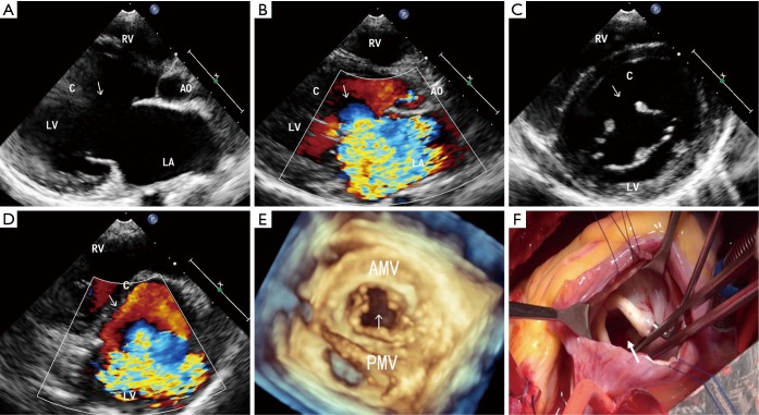
Methods: From October 2012 to June 2016, 19 consecutive patients with MVC diagnosed by 2-dimensional (2D) echocardiography in our hospital were selected for this study. Full-volume RT-3DE was performed on all patients. The 3D-imaging data were cropped and rotated in 3 views (horizontal, sagittal, and coronal) with 6 directions to observe the position and shape of the MVC and the spatial position between the cleft and its surrounding structures. The maximum longitudinal diameter and the maximum width of the cleft were measured. The origin of the mitral regurgitant jet and the severity of MR were evaluated, and these RT-3DE data were compared with the intraoperative findings.
Results: Of the 19 patients studied, 4 patients had isolated cleft mitral valve, and cleft mitral valves combined with other congenital heart lesions were detected in 15 patients. The clefts of 6 patients were located in the A2 segment, the clefts of 4 patients were located in the A1 segment, the clefts of 4 patients were located in the A3 segment, the clefts of 4 patients were located in the A2-A3 segment, and the cleft of 1 patient was located in the P2 segment. Regarding the shape of the cleft, 13 patients had V-shaped clefts, and the others had C- or S-shaped clefts. The severity of the MR at presentation was mild in 2 patients, moderate in 9 and severe in 8. Two of the patients with mild MR did not undergo surgery, while the remaining 17 patients did undergo surgery. Surgical treatment involved direct suture in 11 cases, reconstruction with ring annuloplasty in 3 cases and replacement in 3 cases. The diagnoses of MVC were confirmed by intraoperative findings. RT-3DE successfully captured full-volume 3D images of the 19 patients, which directly displayed the 3D structure of MVC with multiple views such as the position, shape, longitudinal diameter and width of the MVC, and the spatial position between the chordae tendineae surrounding the MVC and the aortic valve. The maximum longitudinal diameter of the valve leaflet cleft measured by RT-3DE and direct measurements during surgery were 12.02±2.12 and 13.01±2.45 mm, respectively, and the difference between these measurements was not statistically significant (P>0.05). Our results indicate that RT-3DE can provide more direct, accurate and abundant information.
Conclusions: RT-3DE is a simple and fast imaging technique, and the detailed 3D images obtained can be used to confirm the diagnosis of MVC. RT-3DE is considered to be an important preoperative test that provides more comprehensive information for selecting a subsequent procedure for treatment.
Keywords: 3-dimensional (3D); Echocardiography; mitral valve cleft (MVC).
Conflict of interest statement
Conflicts of Interest: The authors have no conflicts of interest to declare.
Figures
Similar articles
-
Diagnosis of Isolated Cleft Mitral Valve Using Three-Dimensional Echocardiography.J Am Soc Echocardiogr. 2018 Nov;31(11):1161-1167. doi: 10.1016/j.echo.2018.06.008. Epub 2018 Aug 7. J Am Soc Echocardiogr. 2018. PMID: 30097300 Free PMC article.
-
Revealing Mitral Valve Cleft Using Real-Time 3-Dimensional Echocardiography in Children with Mitral Regurgitation.Pediatr Cardiol. 2024 Mar;45(3):660-665. doi: 10.1007/s00246-023-03155-4. Epub 2023 Apr 5. Pediatr Cardiol. 2024. PMID: 37020140
-
Isolated cleft in the posterior mitral valve leaflet: a congenital form of mitral regurgitation.Clin Cardiol. 2009 Oct;32(10):553-60. doi: 10.1002/clc.20608. Clin Cardiol. 2009. PMID: 19911346 Free PMC article.
-
The Use of 3D Echocardiography in Surgical Planning of the Mitral Valve in Pediatric Cardiology.J Vis Exp. 2021 Jun 3;(172). doi: 10.3791/62574. J Vis Exp. 2021. PMID: 34152324 Review.
-
European Association of Cardiovascular Imaging/Cardiovascular Imaging Department of the Brazilian Society of Cardiology recommendations for the use of cardiac imaging to assess and follow patients after heart transplantation.Eur Heart J Cardiovasc Imaging. 2015 Sep;16(9):919-48. doi: 10.1093/ehjci/jev139. Epub 2015 Jul 2. Eur Heart J Cardiovasc Imaging. 2015. PMID: 26139361 Review.
Cited by
-
Diagnosis of Isolated Cleft of the Anterior Mitral Leaflet in a Dog: A Case Study Using Real-Time Three-Dimensional Echocardiography.Case Rep Vet Med. 2021 Jan 26;2021:6610526. doi: 10.1155/2021/6610526. eCollection 2021. Case Rep Vet Med. 2021. PMID: 33575059 Free PMC article.
-
3D and 4D Ultrasound: Current Progress and Future Perspectives.Curr Cardiovasc Imaging Rep. 2017;10(12):43. doi: 10.1007/s12410-017-9440-2. Epub 2017 Nov 10. Curr Cardiovasc Imaging Rep. 2017. PMID: 29201268 Free PMC article. Review.
-
Diagnosis of Isolated Cleft Mitral Valve Using Three-Dimensional Echocardiography.J Am Soc Echocardiogr. 2018 Nov;31(11):1161-1167. doi: 10.1016/j.echo.2018.06.008. Epub 2018 Aug 7. J Am Soc Echocardiogr. 2018. PMID: 30097300 Free PMC article.
-
Isolated Posterior Mitral Valve Cleft Diagnosed by 3D Echocardiography in an Elderly Woman.JACC Case Rep. 2025 May 21;30(11):103367. doi: 10.1016/j.jaccas.2025.103367. Epub 2025 Apr 23. JACC Case Rep. 2025. PMID: 40409848 Free PMC article.
-
3D echocardiography in mitral valve prolapse.Front Cardiovasc Med. 2023 Jan 10;9:1050476. doi: 10.3389/fcvm.2022.1050476. eCollection 2022. Front Cardiovasc Med. 2023. PMID: 36704460 Free PMC article. Review.
References
LinkOut - more resources
Full Text Sources
Other Literature Sources