

A novel technique for chest drain removal using a two layer method with triclosan-coated sutures
- PMID: 28203426
- PMCID: PMC5303081
- DOI: 10.21037/jtd.2017.01.31
A novel technique for chest drain removal using a two layer method with triclosan-coated sutures
Abstract
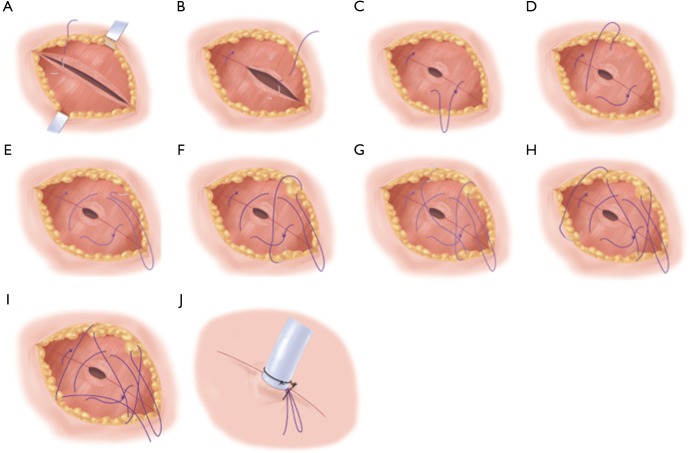
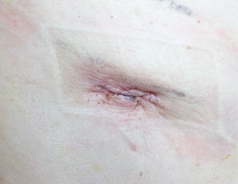
In thoracic surgery, a thoracic drain is always inserted after the surgical procedure. Repair of the wound after removal of the thoracic tube is performed postoperatively, but no universally standard methods currently exists for this tube removal. Here we report a technique using triclosan-coated sutures that is used in thoracic surgery in our hospital. There are several advantages of this technique. First, there is no need for stitches removal on follow-up. Second, it prevents the leakage of pleural exudate because of the tight two-layer sutures. In addition, it was observed to be superior in terms of both wound healing and cosmetic aspects, due to the layer-to-layer sutures. The use of triclosan-coated sutures helps prevent infection and empyema is quite unlikely to occur as the result of the tight ligating of the muscular layer using these sutures. We applied this method in 168 patients over a period of 24 months. There were no complications on removal of the chest tube such as infection, fluid leakage or opening of the surgical wound.
Keywords: New method; thoracic drain; triclosan-coated suture; wound infection.
Conflict of interest statement
Conflicts of Interest: The authors have no conflicts of interest to declare.
Figures
Similar articles
-
Triclosan-coated sutures reduce wound infections after spinal surgery: a retrospective, nonrandomized, clinical study.Spine J. 2015 May 1;15(5):933-8. doi: 10.1016/j.spinee.2013.06.046. Epub 2013 Aug 27. Spine J. 2015. PMID: 23992939
-
The Requirement of Sutures to Close Intercostal Drains Site Wounds in Thoracic Surgery.Ann Thorac Surg. 2018 Feb;105(2):438-440. doi: 10.1016/j.athoracsur.2017.09.032. Epub 2017 Dec 7. Ann Thorac Surg. 2018. PMID: 29223423
-
Knotless suture and hydrocolloid method improves chest drain wound complication.Asian Cardiovasc Thorac Ann. 2022 Sep;30(7):807-812. doi: 10.1177/02184923221106770. Epub 2022 Jun 7. Asian Cardiovasc Thorac Ann. 2022. PMID: 35673271
-
Efficacy of triclosan-coated sutures for reducing risk of surgical site infection in adults: a meta-analysis of randomized clinical trials.J Surg Res. 2016 Mar;201(1):105-17. doi: 10.1016/j.jss.2015.10.015. Epub 2015 Oct 23. J Surg Res. 2016. PMID: 26850191 Review.
-
Triclosan-coated sutures for the prevention of surgical-site infections: a meta-analysis.Acta Chir Belg. 2017 Jun;117(3):137-148. doi: 10.1080/00015458.2017.1287396. Epub 2017 Feb 9. Acta Chir Belg. 2017. PMID: 28399780 Review.
Cited by
-
Pre-embedding subcutaneous suture for chest-tube insertion in uniportal video-assisted thoracoscopic surgery.J Thorac Dis. 2017 Oct;9(10):E938-E940. doi: 10.21037/jtd.2017.09.111. J Thorac Dis. 2017. PMID: 29268440 Free PMC article. No abstract available.
-
An improved method of anchoring chest drain and suture technique for Uni-portal VATS incision.Gen Thorac Cardiovasc Surg. 2021 Nov;69(11):1515-1518. doi: 10.1007/s11748-021-01699-x. Epub 2021 Sep 13. Gen Thorac Cardiovasc Surg. 2021. PMID: 34515949 Free PMC article.
-
Drainology: Leveraging research in chest-drain management to enhance recovery after cardiothoracic surgery.JTCVS Tech. 2024 Apr 9;25:226-240. doi: 10.1016/j.xjtc.2024.04.001. eCollection 2024 Jun. JTCVS Tech. 2024. PMID: 38899104 Free PMC article. No abstract available.
-
Evaluation of the efficacy of a modified method of treating the incisions of the single-port video-assisted thoracoscopic surgery using V-Loc™ barbed sutures.Int Wound J. 2023 Oct;20(8):3131-3139. doi: 10.1111/iwj.14189. Epub 2023 May 4. Int Wound J. 2023. PMID: 37143445 Free PMC article.
-
Feasibility and Safety of a New Chest Drain Wound Closure Method with Knotless Sutures.Korean J Thorac Cardiovasc Surg. 2018 Aug;51(4):260-265. doi: 10.5090/kjtcs.2018.51.4.260. Epub 2018 Aug 5. Korean J Thorac Cardiovasc Surg. 2018. PMID: 30109204 Free PMC article.
References
LinkOut - more resources
Full Text Sources
Other Literature Sources
Medical