

Minimally invasive surgery for giant esophageal leiomyoma: a case report & review of the literatures
- PMID: 28203434
- PMCID: PMC5303089
- DOI: 10.21037/jtd.2017.01.34
Minimally invasive surgery for giant esophageal leiomyoma: a case report & review of the literatures
Abstract
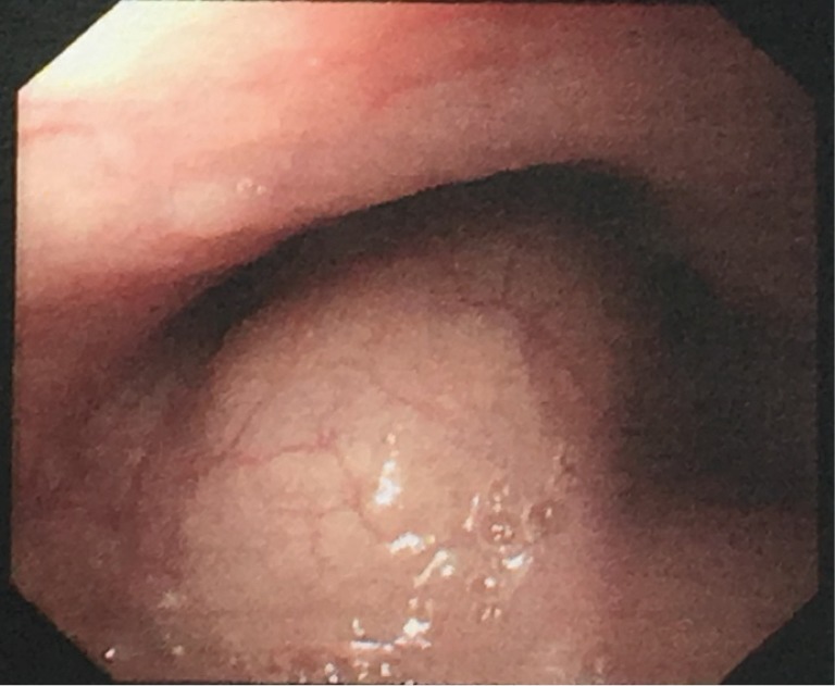
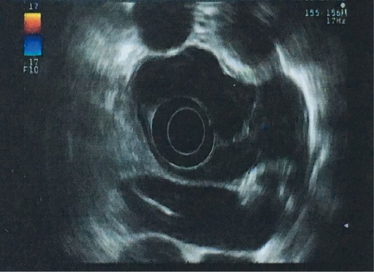
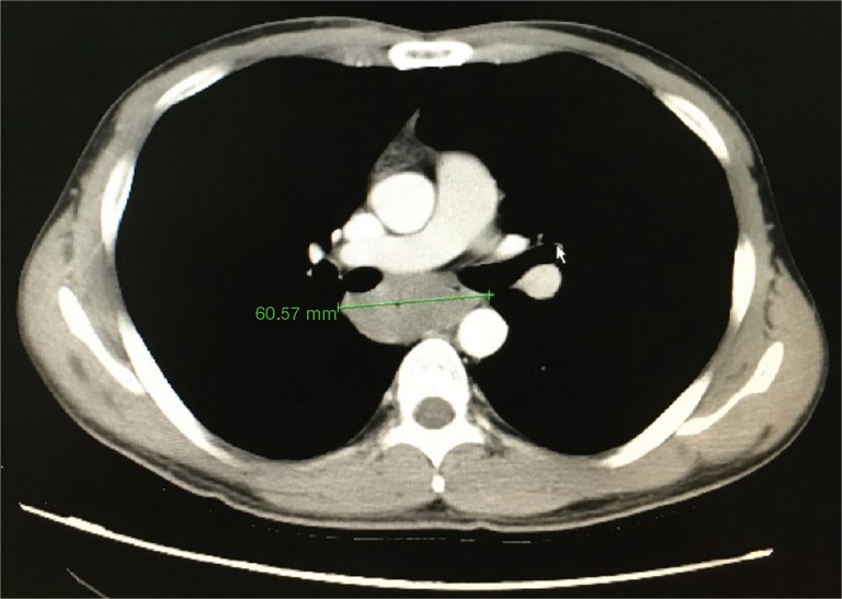
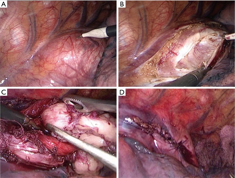
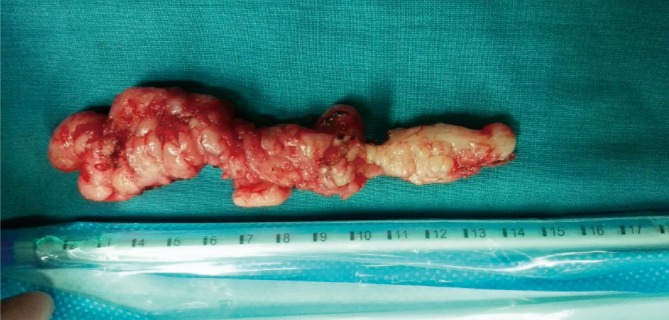
Despite the rapid development of minimally invasive surgery, the treatment of esophageal lesions remains controversial. Giant esophageal leiomyoma could be removed once diagnosed, but its operative method is not quite the same as esophageal leiomyoma of small size. We report a case of giant esophageal leiomyoma and review published cases of giant leiomyomas in the past 10 years. A 29-year-old man was admitted to the clinic for the complaints of 2-month history of dysphagia and discomfort. Radiologic and endoscopic findings suggested esophageal lesion in the muscular layer. The VATS enucleation was performed to relieve the patient's symptoms. The patient started oral intake on the 1st postoperative day, with following solid meal. The postoperative course was uneventful, and the patient was discharged on the 8th postoperative day.
Keywords: Giant esophageal leiomyoma; VATS; enucleation; minimally invasive surgery.
Conflict of interest statement
Conflicts of Interest: The authors have no conflicts of interest to declare.
Figures
Similar articles
-
[Leiomyoma of the esophagus : A further indication for robotic surgery?].Chirurg. 2019 Feb;90(2):125-130. doi: 10.1007/s00104-019-0792-9. Chirurg. 2019. PMID: 30666360 Review. German.
-
Video Assisted Thoracoscopic Surgical Enucleation of a Giant Esophageal Leiomyoma Presenting with Persistent Cough.Case Rep Surg. 2016;2016:7453259. doi: 10.1155/2016/7453259. Epub 2016 Feb 9. Case Rep Surg. 2016. PMID: 26977331 Free PMC article.
-
Robot assisted thoracoscopic resection of giant esophageal leiomyoma.Int J Surg Case Rep. 2014;5(12):1132-4. doi: 10.1016/j.ijscr.2014.11.003. Epub 2014 Nov 11. Int J Surg Case Rep. 2014. PMID: 25460487 Free PMC article.
-
[Enucleation for a giant esophageal leiomyoma; report of a case].Kyobu Geka. 2004 Dec;57(13):1245-9. Kyobu Geka. 2004. PMID: 15609667 Japanese.
-
Robotic-assisted enucleation of a large lower esophageal leiomyoma and review of literature.Int J Med Robot. 2013 Sep;9(3):253-7. doi: 10.1002/rcs.1484. Epub 2013 Feb 12. Int J Med Robot. 2013. PMID: 23401224 Review.
Cited by
-
Laparoscopic approach in the treatment of large leiomyoma of the lower third of the esophagus.Wideochir Inne Tech Maloinwazyjne. 2017 Dec;12(4):437-442. doi: 10.5114/wiitm.2017.72327. Epub 2017 Dec 29. Wideochir Inne Tech Maloinwazyjne. 2017. PMID: 29362660 Free PMC article.
-
Robot-assisted thoracoscopic enucleation for a large esophageal leiomyoma: a case report.Surg Case Rep. 2021 May 26;7(1):129. doi: 10.1186/s40792-021-01212-9. Surg Case Rep. 2021. PMID: 34037886 Free PMC article.
-
Video-assisted thoracoscopic surgery (VATS) enucleation of large gastroesophageal junction leiomyoma: A case report.Int J Surg Case Rep. 2022 Sep;98:107564. doi: 10.1016/j.ijscr.2022.107564. Epub 2022 Aug 31. Int J Surg Case Rep. 2022. PMID: 36058160 Free PMC article.
-
Clinical outcomes in the surgical treatment of esophageal leiomyoma: A retrospective evaluation of 13 cases.Turk Gogus Kalp Damar Cerrahisi Derg. 2020 Jul 28;28(3):505-513. doi: 10.5606/tgkdc.dergisi.2020.18889. eCollection 2020 Jul. Turk Gogus Kalp Damar Cerrahisi Derg. 2020. PMID: 32953214 Free PMC article.
-
Life-threatening giant esophageal neurofibroma with severe tracheal stenosis: a case report.Surg Case Rep. 2018 Sep 3;4(1):107. doi: 10.1186/s40792-018-0517-1. Surg Case Rep. 2018. PMID: 30178113 Free PMC article.
References
-
- Karagülle E, Akkaya D, Türk E, et al. Giant leiomyoma of the esophagus: a case report and review of the literature. Turk J Gastroenterol 2008;19:180-3. - PubMed
Publication types
LinkOut - more resources
Full Text Sources
Other Literature Sources
Miscellaneous