Review
. 2017 Jan;9(1):E71-E77.
doi: 10.21037/jtd.2017.01.49.

Tracheobronchial tuberculosis: a clinical review

Affiliations
Review

Tracheobronchial tuberculosis: a clinical review

Wen Ting Siow et al. J Thorac Dis. 2017 Jan.

Abstract

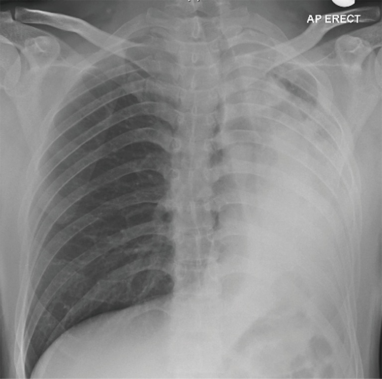
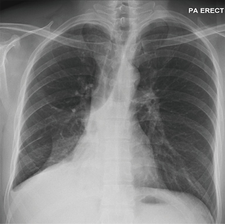
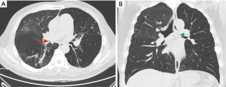
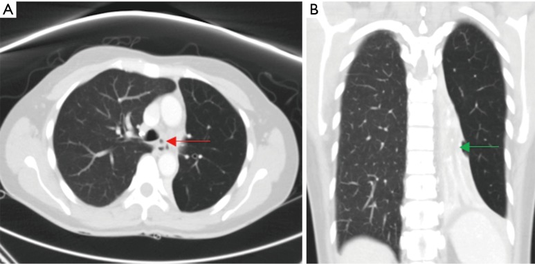
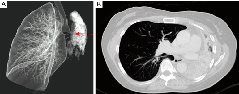
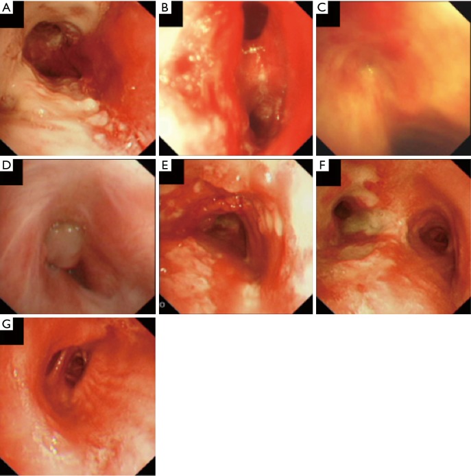
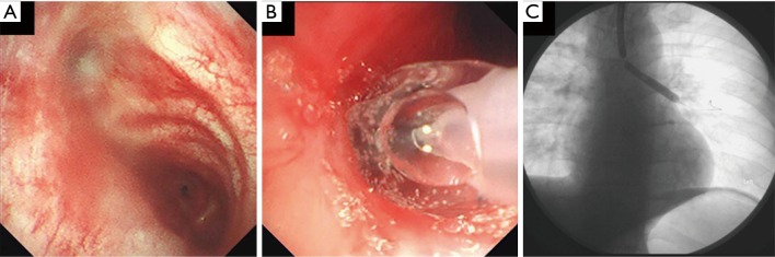
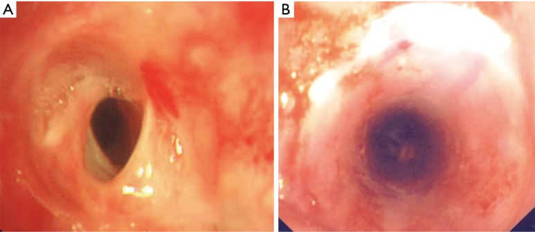
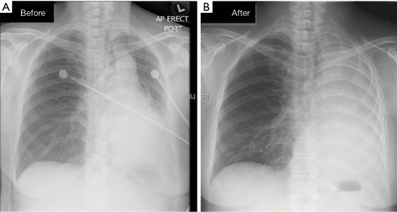
Endobronchial tuberculosis (EBTB) is defined as tuberculous infection of the tracheobronchial tree. The exact pathogenesis is unclear, and it has a heterogenous clinical course. Its diagnosis requires the clinician to have a high index of suspicion based on clinical symptoms and radiological features. Computed tomography and bronchoscopy are useful tools in its evaluation. The goal of treatment is in the eradication of tuberculous bacilli with appropriate anti-tuberculous therapy. Use of corticosteroids is controversial for the prevention of tracheobronchial stenosis. Interventional bronchoscopy or surgical intervention is employed to restore airway patency once significant stenosis occurs.

Keywords: Endobronchial; bronchoscopy; stenosis; tracheobronchial; tuberculosis (TB).


Conflict of interest statement

Conflicts of Interest: The authors have no conflicts of interest to declare.

Figures

Figure 1
Figure 1
Chest roentography showing atelectasis of left lung with partial aeration of left upper lobe.
Figure 2
Figure 2
Right bronchus intermedius stenosis resulting in middle and lower lobe collapse.
Figure 3
Figure 3
CT thorax showing left main bronchus (red arrow) and right bronchus intermedius stenosis (green arrow), with distal tree-in-bud infiltrates.
Figure 4
Figure 4
CT Thorax showing left main bronchus narrowing (red arrow) and resultant distal atelectasis (green arrow).
Figure 5
Figure 5
3D-reconstruction of airways showing stricture of left main bronchus (red arrow) and resultant distal atelectasis.
Figure 6
Figure 6
Bronchoscopic appearances of endobronchial TB. (A) Actively caseating; (B) oedematous-hyperaemic; (C) fibrostenotic subtype; (D) tumorous subtype; (E) granular subtype; (F) ulcerative subtype; (G) nonspecific bronchitic subtype.
Figure 7
Figure 7
Balloon dilation of left main bronchus stricture. (A) Stricture before dilation; (B) balloon dilation in progress; (C) fluoroscopic view of balloon and guidewire in left main bronchus.
Figure 8
Figure 8
Fibro-stenotic left main stricture (A), radial cuts applied with electrosurgical knife, dilated with balloon bronchoplasty (not demonstrated), and silicon stent placement (B).
Figure 9
Figure 9
Before and after left pneumonectomy for post tuberculous endobronchial stricture, after failing balloon dilatation of left main bronchial stricture.

References

    1. World Health Organisation. Global tuberculosis report 2015. Available online: http://apps.who.int/iris/bitstream/10665/191102/1/9789241565059_eng.pdf
    1. Smart J. Endo-bronchial tuberculosis. Br J Tuberc Dis Chest 1951;45:61-8. 10.1016/S0366-0869(51)80003-0 - DOI - PubMed
    1. Lee JH, Park SS, Lee DH, et al. Endobronchial Tuberculosis: Clinical and Bronchoscopic Features in 121 Cases. Chest 1992;102:990-4. 10.1378/chest.102.4.990 - DOI - PubMed
    1. Jung SS, Park HS, Kim JO, et al. Incidence and clinical predictors of endobronchial tuberculosis in patients with pulmonary tuberculosis. Respirology 2015;20:488-95. 10.1111/resp.12474 - DOI - PMC - PubMed
    1. Van den Brande PM, Van de Mierop F, Verbeken EK, et al. Clinical Spectrum of Endobronchial Tuberculosis in Elderly Patients. Arch Intern Med 1990;150:2105-8. 10.1001/archinte.1990.00390210081018 - DOI - PubMed

LinkOut - more resources