

Complex chromosomal rearrangements by single catastrophic pathogenesis in NUT midline carcinoma
- PMID: 28203693
- PMCID: PMC5378225
- DOI: 10.1093/annonc/mdw686
Complex chromosomal rearrangements by single catastrophic pathogenesis in NUT midline carcinoma
Abstract
Background: Nuclear protein in testis (NUT) midline carcinoma (NMC) is a rare aggressive malignancy often occurring in the tissues of midline anatomical structures. Except for the pathognomonic BRD3/4-NUT rearrangement, the comprehensive landscape of genomic alterations in NMCs has been unexplored.
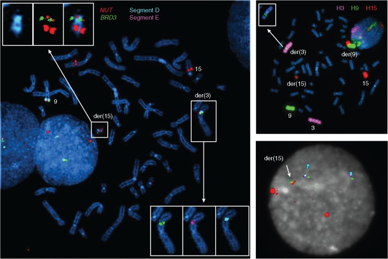
Patients and methods: We investigated three NMC cases, including two newly diagnosed NMC patients in Seoul National University Hospital, and a previously reported cell line (Ty-82). Whole-genome and transcriptome sequencing were carried out for these cases, and findings were validated by multiplex fluorescence in situ hybridization and using individual fluorescence probes.
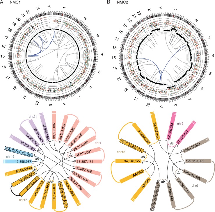
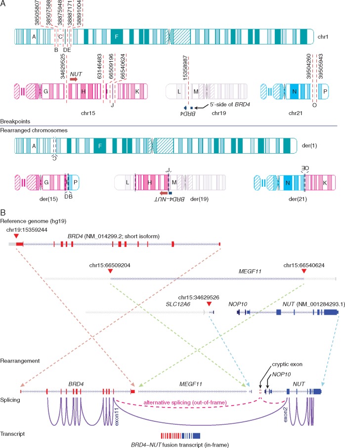
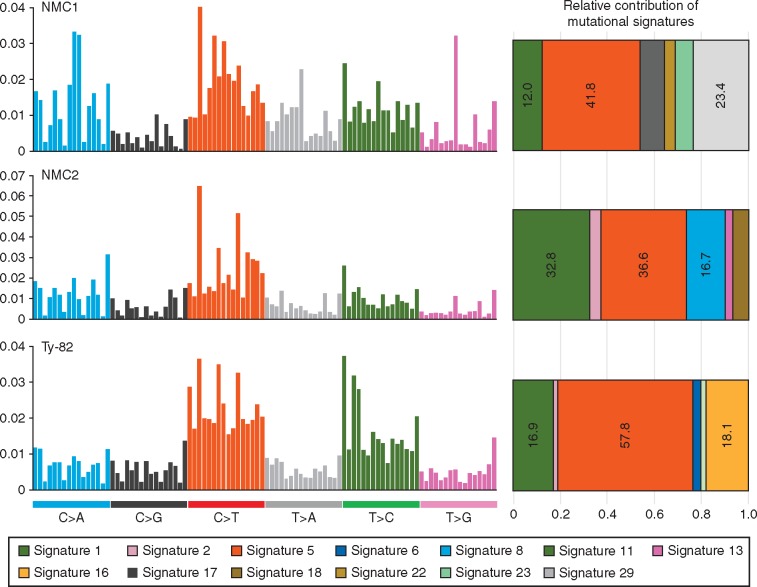
Results: Here, we present the first integrative analysis of whole-genome sequencing, transcriptome sequencing and cytogenetic characterization of NUT midline carcinomas. By whole-genome sequencing, we identified a remarkably similar pattern of highly complex genomic rearrangements (previously denominated as chromoplexy) involving the BRD3/4-NUT oncogenic rearrangements in two newly diagnosed NMC cases. Transcriptome sequencing revealed that these complex rearrangements were transcribed as very simple BRD3/4-NUT fusion transcripts. In Ty-82 cells, we also identified a complex genomic rearrangement involving the BRD4-NUT rearrangement underlying the simple t(15;19) karyotype. Careful inspections of rearrangement breakpoints indicated that these rearrangements were likely attributable to single catastrophic events. Although the NMC genomes had >3000 somatic point mutations, canonical oncogenes or tumor suppressor genes were rarely affected, indicating that they were largely passenger events. Mutational signature analysis showed predominant molecular clock-like signatures in all three cases (accounting for 54%-75% of all base substitutions), suggesting that NMCs may arise from actively proliferating normal cells.
Conclusion: Taken together, our findings suggest that a single catastrophic event in proliferating normal cells could be sufficient for neoplastic transformation into NMCs.
Keywords: NUT midline carcinoma; bromodomain and extra terminal; chromoplexy; complex genomic rearrangement; mutational signature.
© The Author 2017. Published by Oxford University Press on behalf of the European Society for Medical Oncology.
Figures

References
-
- Kees UR, Mulcahy MT, Willoughby ML.. Intrathoracic carcinoma in an 11-year-old girl showing a translocation t(15;19). Am J Pediatr Hematol Oncol 1991; 13: 459–464. - PubMed
-
- Kubonishi I, Takehara N, Iwata J. et al. Novel t(15;19)(q15;p13) chromosome abnormality in a thymic carcinoma. Cancer Res 1991; 51: 3327–3328. - PubMed
-
- French CA. Pathogenesis of NUT midline carcinoma. Annu Rev Pathol 2012; 7: 247–265. - PubMed
-
- Travis WD, Brambilla E, Nicholson AG. et al. The 2015 World Health Organization classification of lung tumors: impact of genetic, clinical and radiologic advances since the 2004 classification. J Thorac Oncol 2015; 10: 1243–1260. - PubMed
Publication types
MeSH terms
Substances
LinkOut - more resources
Full Text Sources
Other Literature Sources
Molecular Biology Databases
Research Materials
