. 2017 Feb 1;96(2):352-365.
doi: 10.1095/biolreprod.116.143735.

Redistribution of soluble N-ethylmaleimide-sensitive-factor attachment protein receptors in mouse sperm membranes prior to the acrosome reaction

Affiliations

Redistribution of soluble N-ethylmaleimide-sensitive-factor attachment protein receptors in mouse sperm membranes prior to the acrosome reaction

Momal Sharif et al. Biol Reprod. .

Abstract

Formation of complexes between soluble N-ethylmaleimide-sensitive-factor attachment protein receptor (SNARE) proteins on opposing membranes is the minimal requirement for intracellular membrane fusion. The SNARE, syntaxin 2, is found on the sperm plasma membrane and a second SNARE, vesicle associated membrane protein 2 (VAMP2, also known as synaptobrevin 2, SYB2), is on the apposing outer acrosomal membrane. During the acrosome reaction, the outer acrosomal membrane fuses at hundreds of points with the plasma membrane. We hypothesized that syntaxin 2 and VAMP2 redistribute within their respective membranes prior to the acrosome reaction to form trans-SNARE complexes and promote membrane fusion. Immunofluorescence and superresolution structured illumination microscopy were used to localize syntaxin 2 and VAMP2 in mouse sperm during capacitation. Initially, syntaxin 2 was found in puncta throughout the acrosomal region. At 60 and 120 min of capacitation, syntaxin 2 was localized in puncta primarily in the apical ridge. Although deletion of bicarbonate during incubation had no effect, syntaxin 2 puncta were relocated in the restricted region in less than 20% of sperm incubated without albumin. In contrast, VAMP2 was already found in puncta within the apical ridge prior to capacitation. The puncta containing syntaxin 2 and VAMP2 did not precisely co-localize at 0 or 60 min of capacitation time. In summary, syntaxin 2 shifted its location to the apical ridge on the plasma membrane during capacitation in an albumin-dependent manner but VAMP2 was already localized to the apical ridge. Puncta containing VAMP2 did not co-localize with those containing syntaxin 2 during capacitation; therefore, formation of trans-SNARE complexes containing these SNAREs does not occur until after capacitation, immediately prior to acrosomal exocytosis.

Keywords: SNAREs; sperm; acrosome reaction; exocytosis; capacitation; syntaxin 2 (STX2); vesicle associated membrane protein 2 (VAMP2); membrane fusion; plasma membrane.


Figures

Figure 1.
Figure 1.
Model of SNARE function in membrane fusion leading to completion of the acrosome reacion. (A) Acrosome intact mouse sperm with plasma membrane (PM), outer acrosomal membrane (OAM), and inner acrosomal membrane (IAM) immediately prior to membrane interaction. Syntaxin 2 (STX2) and SNAP25 are present on the plasma membrane, while VAMP2 is apposed to them on the outer acrosomal membrane. (B) Trans-SNARE complex formation is promoting interaction of the plasma membrane and outer acrosomal membrane. (C) Membrane fusion is leading to formation of fusion pores, leading to release of acrosomal contents. (D) Sperm after the release of the plasma and outer acrosomal membranes.
Figure 2.
Figure 2.
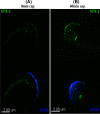
Location of syntaxin 2 (STX2) in capacitated and noncapacitated sperm using confocal microscopy. Sperm were collected and incubated under noncapacitating (ncap, A) and capacitating (cap, B) conditions for 60 min. Sperm were fixed, permeabilized, and STX2 (green) was detected with an antibody. (A) Sperm that were incubated in noncapacitating medium displayed STX2 in a punctate pattern over much of the sperm head. (B) Sperm that were incubated in capacitating medium displayed STX2 mostly at the apical ridge of the sperm head. (C) If sperm were incubated with nonimmune IgG rather than syntaxin antibody, no staining was observed.
Figure 3.
Figure 3.
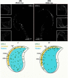
SR-SIM detects syntaxin 2 (STX2) change in distribution in sperm during capacitation. Sperm were collected and incubated under capacitating conditions for 60 min. Sperm were fixed, permeabilized, and STX2 was detected with an antibody and SR-SIM. (A) Images of sperm incubated in noncapacitating medium (ncap). A punctate pattern of STX2 was observed scattered over the sperm head in the large image as well as three insets. (A’) Schematic representation of (A). (B) Images of sperm incubated in capacitating media (cap). Puncta-containing STX2 were observed primarily at the apical ridge of the sperm head. This is observed in the large image and three insets. (B’) Schematic representation of (B).
Figure 4.
Figure 4.
Syntaxin 2 (STX2) change in location is not due to acrosomal exocytosis. Sperm were stained with antibodies to STX2 (green) and ZP3R (blue). Images are of sperm incubated in capacitating media for 0 min (A) or 60 min (B) and examined by SR-SIM. Upper images show STX2 localization and lower images show STX2 and ZP3R overlay. At 60 min, STX2 was localized to the apical ridge in sperm that were stained by the ZP3R antibody and therefore had an intact acrosome.
Figure 5.
Figure 5.
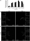
The location of syntaxin 2 (STX2) in sperm incubated in albumin- or bicarbonate-free medium. (A) The pattern of STX2 localization was analyzed from the images collected at different time points (0, 10, 30, 60, or 120 min) from sperm incubated in capacitating medium (C), medium without BSA (-A), and medium without HCO3 (–H). (B) Sperm incubated in medium with no HCO3 for 0 min showed punctate STX2 localization over the sperm head in the large as well as three insets. Sperm incubated in media with no HCO3 for 60 min showed punctate labeling more abundantly at the apical ridge of the sperm head in the large image and three insets. (C) Sperm incubated in medium with no albumin for 0 min showed punctate STX2 localization over the sperm head in the large as well as three insets. Sperm incubated in media with no albumin for 60 min showed punctate labeling scattered over the sperm head in the large image and three insets, much like sperm at 0 min. A total of 16 sperm were analyzed for each treatment and time point.
Figure 6.
Figure 6.
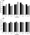
The change in location of syntaxin 2 (STX2) during capacitation does not change the average size of STX2 positive puncta and STX2 positive volume. (A) Quantification of images collected using SR-SIM. The average size of STX2 positive puncta on sperm head was calculated at different time points (0, 10, 30, 60, or 120 min) from sperm incubated in capacitating medium (C), media without BSA (–A), and medium without HCO3 (–H). A total of 16 sperm were analyzed for each treatment and time point. No statistically significant difference was found. (B) The STX2 positive fluorescence volume on the sperm head was calculated. Images were collected at 0, 10, 30, 60, or 120 min from sperm incubated in capacitating medium (C), medium without BSA (-A), and medium without HCO3 (-H). A total of 16 sperm were analyzed for each treatment and time point. No statistically significant difference was observed.
Figure 7.
Figure 7.
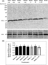
Relocalization of syntaxin 2 (STX2) during capacitation does not change the abundance of STX2. Sperm were incubated for 0, 30, 60, or 120 min in capacitating media or for 60 min in medium without BSA (–A), without HCO3 (–H), and without both HCO3 and BSA (–A/–H). Protein lysates were characterized by immunoblotting with a STX2 antibody. Actin was used as a loading control and mouse brain was used as a positive control. (A) Syntaxin 2 immunoblotting. There was no change in the amount of STX2 collected from mouse sperm. (B) Graphical representation of the STX2 immunoblotting data. Syntaxin 2 was normalized against actin band intensities. No significant difference was observed; n = 3.
Figure 8.
Figure 8.
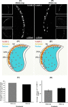
VAMP2 detected by SR-SIM does not redistribute in sperm during capacitation. (A) Images of sperm incubated in noncapacitating medium (ncap) for 60 min. A punctate pattern of VAMP2 can be observed at the apical ridge of the sperm in the large image as well as three insets. () Schematic representation of (A). (B) Images of sperm incubated in capacitating medium (cap) for 60 min. A similar pattern of VAMP2 can be observed at the apical ridge of the sperm head when compared with (A). () Schematic representation of (B). The larger image and three insets show a similar result. (C) Images sperm at 60 in of incubation in capacitating medium and noncapacitating conditions collected using SR-SIM microscopy were quantified. The percentage of sperm with VAMP2 at the apical ridge was plotted. (D) The volume of VAMP2-positive fluorescence on sperm head was calculated. Images were collected at 60 min from sperm incubated in capacitating and noncapacitating medium. A total of 16 sperm were analyzed for each treatment and time point. No statistical difference was found.
Figure 9.
Figure 9.
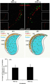
Syntaxin 2 (STX2) and VAMP2 do not co-localize in capacitated sperm. Sperm were incubated for 60 min in capacitating conditions and stained with antibodies and appropriate fluorescent secondary antibodies to STX2 (green) and VAMP2 (red). No co-localization was observed between STX2 and VAMP2 when images collected at either 0 min (A) or 60 min (B) of capacitation time. ( and ) are schematic representations of (A and B). (C) Graphical representation of co-localization data obtained from the SR-SIM images. A total of 16 sperm were analyzed for each treatment and time point. There was no significant correlation in localization observed at either 0 or 60 min sperm capacitation time.

Similar articles

Cited by

References

    1. Salicioni AM, Platt MD, Wertheimer EV, Arcelay E, Allaire A, Sosnik J, Visconti PE. Signaling pathways involved in sperm capacitation. Soc Reprod Fertil Suppl, 2007: 65:245–259. - PubMed
    1. Abou-haila A, Tulsiani DRP. Signal transduction pathways that regulate sperm capacitation and the acrosome reaction. Arch Biochem Biophys, 2009, 485:72–81. - PubMed
    1. De Lamirande E., Capacitation as a regulatory event that primes spermatozoa for the acrosome reaction and fertilization. Mol Hum Reprod, 1997, 3:175–194. - PubMed
    1. Flesch FM, Brouwers JF, Nievelstein PF, Verkleij AJ, van Golde LM, Colenbrander B, Gadella BM. Bicarbonate stimulated phospholipid scrambling induces cholesterol redistribution and enables cholesterol depletion in the sperm plasma membrane. J Cell Sci, 2001, 114:3543–3555. - PubMed
    1. Nolan JP, Hammerstedt RH. Regulation of membrane stability and the acrosome reaction in mammalian sperm. FASEB J, 1997, 11:670–682. - PubMed