. 2017 Feb 28;45(4):2016-2028.
doi: 10.1093/nar/gkw842.

Unusual C΄/D΄ motifs enable box C/D snoRNPs to modify multiple sites in the same rRNA target region

Affiliations

Unusual C΄/D΄ motifs enable box C/D snoRNPs to modify multiple sites in the same rRNA target region

Robert Willem van Nues et al. Nucleic Acids Res. .

Abstract

Eukaryotic box C/D small nucleolar (sno)RNPs catalyse the site-specific 2΄-O-methylation of ribosomal RNA. The RNA component (snoRNA) contains guide regions that base-pair with the target site to select the single nucleotide to be modified. The terminal C/D and internal C΄/D΄ motifs in the snoRNA, adjacent to the guide region, function as binding sites for the snoRNP proteins including the enzymatic subunit fibrillarin/Nop1. Four yeast snoRNAs are unusual in that they are predicted to methylate two nucleotides in a single target region. In each case, the internal C΄/D΄ motifs from these snoRNAs differ from the consensus. Our data indicate that the C΄/D΄ motifs in snR13, snR48 and U18 form two alternative structures that lead to differences in the position of the proteins bound to this motif. We propose that each snoRNA forms two different snoRNPs, subtly different in how the proteins are bound to the C΄/D΄ motif, leading to 2΄-O-methylation of different nucleotides in the target region. For snR48 and U18, the unusual C΄/D΄ alone is enough for the modification of two nucleotides. However, for the snR13 snoRNA the unusual C΄/D΄ motif and extra base-pairing, which stimulates rRNA 2΄-O-methylation, are both critical for multiple modifications in the target region.


Figures
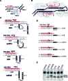
Figure 1.
Figure 1.
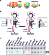
The 2΄-O-methylation activity of snR13, snR48, U18 and U24 C΄/D΄ motifs. (A) Schematic representation of the box C/D motif with the adjacent guide sequence base-paired to the rRNA target (shown in red with the target nucleotide indicated by an asterisk). The sequence of the C and D boxes are shown. The positions of stem I and II in the motif are indicated. Note that the C/D motif is rotated 180° relative to its orientation in (B) so that it is in the same orientation as the C΄/D΄ motif. (B) A schematic model of the box C/D snoRNP complex. The snoRNA is shown in black, with the C, D, C΄ and D΄ boxes indicated. The rRNA is shown in red, with the base-pairing interactions indicated. The asterisk represents the site modified. The box C/D snoRNP proteins are represented as grey filled ellipses. (C) rRNA (upper) and snoRNA (lower) sequences, with both conventional guide-rRNA interactions (rRNA shaded red) and novel extra (or accessory) base-pairing (rRNA shaded blue) interactions, for the S. cerevisiae box C/D snoRNAs that direct multi-site modification. Where sequences are shaded both red and blue, this indicates an overlap between the conventional and extra-base-pairing. The C΄ and D΄ sequences are shown in white with a black background. The guide sequences in the snoRNA are indicated. Note for snR48 there is no obvious D΄ box and the whole conserved area is marked (see later). The modified nucleotides are indicated by an asterisk together with the rRNA nucleotide number. (D) The guide sequence and D΄ motif of the artificial snoRNA constructs used to test the function of the various C΄/D΄ motifs is shown (the full sequence used is shown in Supplementary Figure S1B and C). The D΄ boxes are presented in white with a black background. The target 18S sequence (white text on red background) is shown with the expected (black background) and actual (asterisk) methylation sites indicated. (E) Constructs expressing artificial snoRNAs containing the C΄/D΄ motifs from U18, U24, snR13, snR48 and human (h)U24 (as indicated above each lane) were transformed into yeast cells. RNA was extracted from the cells and analysed by primer extension to detect rRNA methylation. The position of the stop corresponding to methylation of the target nucleotides, S1314 and S1316 in the 18S rRNA, are indicated on the left. Bands corresponding to 2΄-O-methylation are indicated by an asterisk. Northern blotting was used to control for snoRNA expression (Supplementary Figure S2A).
Figure 2.
Figure 2.
The 5΄ end of box D΄ and the extra nucleotide in box C΄ are both required for multiple site 2΄-O-methylation by the U18 snoRNA. (A) A WebLogo representation of the evolutionary conservation of the guide, D΄ box, accessory guide and C΄ box sequences (as indicated below) of the U18 snoRNA derived from the alignment of all the available yeast U18 snoRNA sequences (24). The consensus C΄ and D΄ sequences are shown above. The diagram was prepared using the WebLogo software (29). (B) Two secondary structure models, one showing canonical base-pairing the other an alternative structure for stem II, of the U18mutX box C΄/D΄ motif and guide sequence, in the context of the artificial snoRNA, base-paired to the 18S rRNA. Note, in the alternative structure a longer, 5 nucleotide box D΄ is used. The U18mutX RNA differs from the wild-type U18 in the sequence of the accessory guide region (Supplementary Figure S1C) that has been altered to base-pair with the 18S rRNA adjacent the region targeted by the artificial snoRNA. The arrows indicate the site modified. The box C΄/D΄ motif and the accessory guide are shown in white on a black background. The positions of the C΄, D΄ boxes and stem II are indicated. The sequence of the box C΄ and D΄ mutants is also shown to the right and left of the structures, respectively. The numbering system used starts at the first position of the canonical box sequence. (C) Plasmids expressing artificial snoRNAs containing the wild-type and mutant U18 C΄/D΄ motifs (as indicated above each lane) were transformed into yeast cells. RNA was extracted from the cells and analysed by primer extension to detect rRNA methylation. The position of the stop corresponding to methylation of the target nucleotides, S1315 and S1316 in the 18S rRNA, are indicated. The levels of the various snoRNAs were determined by Northern blotting (Supplementary Figure S2B).
Figure 3.
Figure 3.
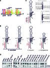
The snR48 C'D' motif is unique and directs 2΄-O-methylation at multiple sites in the target region. (A) A WebLogo representation of the conservation of the guide, D΄ box and C΄ box sequences of the snR48 snoRNA derived from the alignment of all the available yeast snR48 sequences (24). The consensus sequence for the C΄ and D΄ boxes is shown above the WebLogo image. Note, two potential alternative positions for the D΄ box are shown. The diagram was prepared using the WebLogo software (29). (B) Secondary structure of the snR48 box C΄/D΄ motif and guide sequence base-paired to the 25S rRNA from S. cerevisiae, S. kluyveri and L. elongisporus. Note two alternative structures are shown for the S. cerevisiae snR48. The C΄ and D΄ boxes are shown in white on a black background. Arrows indicate the site to be modified. (C) Secondary structure of the snR48 box C΄/D΄ motif, in the context of the artificial snoRNA targeting sites 1314 and 1316 in the 18S rRNA. The C΄ and D΄ boxes are shown in white on a black background. Arrows indicate the site to be modified. The mutations to the C΄ and D΄ boxes are shown to the right. (D) Constructs expressing artificial snoRNAs containing the wild-type and mutant snR48 C΄/D΄ motifs (as indicated above each lane) were transformed into yeast cells. RNA was extracted from the cells and analysed by primer extension to detect rRNA methylation. The position of the two target nucleotides (S1314 and S1316) is indicated on the right. L.elo is the artificial snoRNA with the C΄/D΄ motif from L. elongisporus snR48. snR48 glucose: yeast containing the plasmid encoding the artificial snoRNA with the snR48 C΄/D΄ motif, grown on glucose containing media. The levels of the various snoRNAs were determined by Northern blotting (Supplementary Figure S2C).
Figure 4.
Figure 4.
The last nucleotide of the guide region can influence the site to be 2΄-O-methylated in the target RNA. (A) A schematic representation of the conservation of the active guide and D΄/D box sequences of the S. cerevisiae snoRNAs (24). The diagram was prepared using the WebLogo software (29). (B) Comparison of the 3΄ nucleotide of the active and inactive (no guide) guide regions of the available yeast box C/D snoRNAs (24). (C) Secondary structure of the hU24 box C΄/D΄ motif, in the context of the artificial snoRNA targeting sites 1316 in the 18S rRNA. The C΄ and D΄ boxes are shown in white on a black background. The arrow indicates the site to be modified. The mutations to the D΄ box are shown to the left. (D) Constructs expressing artificial snoRNAs containing the wild-type and mutant hU24 C΄/D΄ motifs (as indicated above each lane) were transformed into yeast cells. RNA was extracted from the cells and analysed by primer extension to detect rRNA methylation. The position of the stop corresponding to methylation of the target nucleotides, S1315 and S1316 in the 18S rRNA, are indicated. The levels of the various snoRNAs were determined by Northern blotting (Supplementary Figure S2D).
Figure 5.
Figure 5.
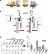
An unusual D΄ box and the extra base-pairing sequence are required for multi-site 2΄-O-methylation by the snR13 snoRNA. (A) A schematic representation of the conservation of the active guide and D΄/D box sequences of the snR13 snoRNA (24).The consensus for the C΄ and D΄ sequences is shown above the image. The diagram was prepared using the WebLogo software (29). (B) Secondary structure models of the snR13 box C΄/D΄ motif, guide region and accessory guide with the natural rRNA target site (shown in red) in the 25S rRNA. The C΄ and D΄ boxes are shown in white on a black background. The arrow indicates the site to be modified. The mutations to the D΄ box are shown to the left and the mutation to the accessory guide is shown on the right. Arrows indicate the site in the rRNA to be modified. Note, in the alternative structure a longer, five nucleotide box D΄ is used. (C) Constructs expressing wild-type and mutant snR13 snoRNAs (as indicated above each lane) were transformed into yeast cells. RNA was extracted from the cells and analysed by primer extension to detect rRNA methylation. The position of the stop corresponding to methylation of the target nucleotides, L2280 and L2281 in the 25S rRNA, are indicated. The levels of the various snoRNAs were determined by Northern blotting (Supplementary Figure S2E). (E) The data presented in (D) was analysed using imagequant software and the relative levels of the bands corresponding to the L2280 and L2281, relative to the signal seen for the modification at L2288, were calculated and plotted. The data were adjusted to the WT signal at L2280. Error bars indicate standard deviation from three separate experiments.
Figure 6.
Figure 6.
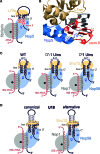
Interaction of Nop56 with stem II of the C΄/D΄ motif dictates the site of 2΄-O-methylation. (A) Schematic model of the Archaeal C΄/D΄ motif and guide region base-paired to a target rRNA (shown in red). The box C/D snoRNP proteins are represented as grey filled ellipses. The black dot indicates the active site of fibrillarin (Nop1 in yeast) and is positioned over the nucleotide to be modified. The Q and R in Nop5 represent amino acids Q296 and R339 that make contact with stem II of both the C/D and C΄/D΄ motif motifs. Stem II is the C΄/D΄ motif is shown in white on a red background. The upper part of the C΄/D΄ motif, which is bound by L7Ae, is shown in white with a black background. (B) Recognition of stem II of the box C/D motif by the Nop domain of Archaeal Nop5 (22). Cartoon views of the relevant regions of Nop5 and L7Ae are shown. Amino acid side chains are only shown for Q296 and R339. The snoRNA is shown in cartoon form and only the C΄/D΄ motif is shown for clarity. Stem II is shown in red and the upper part, bound by L7Ae, is shown in dark grey. The identity of the nucleotides in stem II is indicated. Hydrogen bonds between Q296 and R339 I Nop5 and stem II of the C΄/D΄ motif are shown in yellow. (C) Organisation of the snoRNP proteins on the artificial snoRNA containing the hU24 C΄/D΄ motif and the D΄ box insertions presented in Figure 4. The schematic organisation is the same as in (A) except the whole C΄/D΄ motif is shown in white with a black background. Note the slight, one nucleotide shift, in the position of Nop56, and he corresponding change in Nop1 position, in the complex based on the recognition of stem II in the C΄/D΄ motif. (D) Organisation of the snoRNP proteins on the U18 snoRNA based on the canonical and alternative secondary structures presented in Figure 2B. The schematic organisation is the same as in (C).

Similar articles

Cited by

References

    1. Decatur W.A., Fournier M.J.. rRNA modifications and ribosome function. Trends Biochem. Sci. 2002; 27:344–351. - PubMed
    1. Helm M. Post-transcriptional nucleotide modification and alternative folding of RNA. Nucleic Acids Res. 2006; 34:721–733. - PMC - PubMed
    1. Fischer N., Neumann P., Konevega A.L., Bock L.V., Ficner R., Rodnina M.V., Stark H.. Structure of the E. coli ribosome-EF-Tu complex at <3 A resolution by C-corrected cryo-EM. Nature. 2015; 520:567–570. - PubMed
    1. Motorin Y., Helm M.. RNA nucleotide methylation. Wiley Interdiscip. Rev. RNA. 2011; 2:611–631. - PubMed
    1. Baudin-Baillieu A., Fabret C., Liang X.H., Piekna-Przybylska D., Fournier M.J., Rousset J.P.. Nucleotide modifications in three functionally important regions of the Saccharomyces cerevisiae ribosome affect translation accuracy. Nucleic Acids Res. 2009; 37:7665–7677. - PMC - PubMed

Publication types

LinkOut - more resources