Review
. 2017 Jul;10(4):719-734.
doi: 10.1111/1751-7915.12693. Epub 2017 Feb 16.

Should the biofilm mode of life be taken into consideration for microbial biocontrol agents?

Affiliations
Review

Should the biofilm mode of life be taken into consideration for microbial biocontrol agents?

Caroline Pandin et al. Microb Biotechnol. 2017 Jul.

Abstract

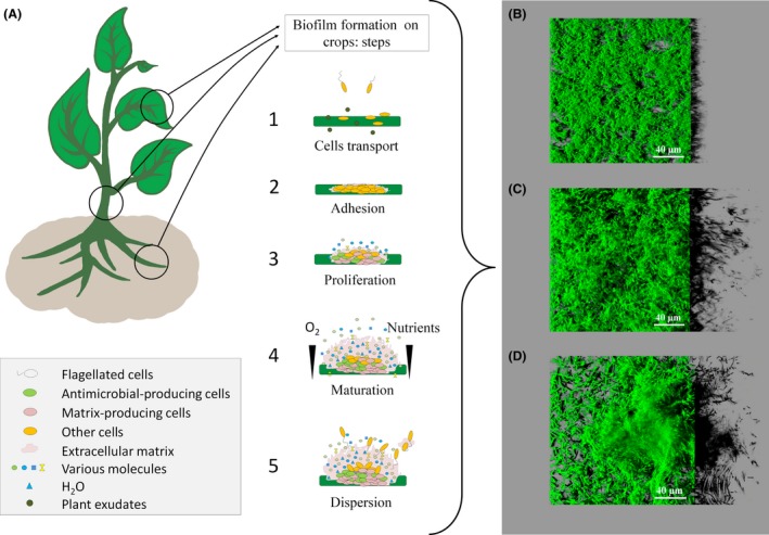
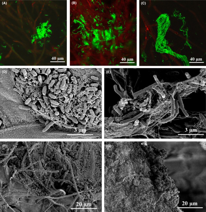
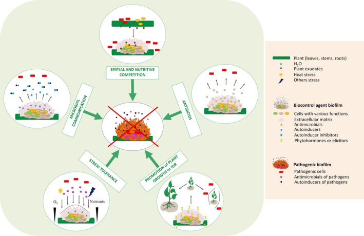
Almost one-third of crop yields are lost every year due to microbial alterations and diseases. The main control strategy to limit these losses is the use of an array of chemicals active against spoilage and unwanted pathogenic microorganisms. Their massive use has led to extensive environmental pollution, human poisoning and a variety of diseases. An emerging alternative to this chemical approach is the use of microbial biocontrol agents. Biopesticides have been used with success in several fields, but a better understanding of their mode of action is necessary to better control their activity and increase their use. Very few studies have considered that biofilms are the preferred mode of life of microorganisms in the target agricultural biotopes. Increasing evidence shows that the spatial organization of microbial communities on crop surfaces may drive important bioprotection mechanisms. The aim of this review is to summarize the evidence of biofilm formation by biocontrol agents on crops and discuss how this surface-associated mode of life may influence their biology and interactions with other microorganisms and the host and, finally, their overall beneficial activity.


Figures

Figure 1
Figure 1
Biofilm formation on crops and in vitro: (A): On crops: The first step involves deposition on the substratum (1) followed by adhesion (2) to the support through cell wall decorations and extracellular appendages. Once attached, a proliferation phase (3) and the diversification of cell types initiate the spatial organization of the biostructure, leading to biofilm maturation (4). Biofilm ageing or environmental conditions unfavourable for the maintenance of the biofilm results in regulated dispersion of the biofilm (5), disseminating free cells and cell clusters that will start a new biofilm cycle on a new surface. B–D. In vitro: Structural diversity of three biocontrol agents as observed in vitro (24 h of axenic culture in microplates at 25°C) by confocal laser scanning microscopy (Leica SP8); (B) Bacillus amyloliquefaciens FZB42 expressing a green fluorescent protein (GFP), forming flat undifferentiated architecture, (C) Bacillus amyloliquefaciens SQR9 expressing a GFP and (D) Bacillus subtilis QST 713 (labelled in green with syto 9, Invitrogen, France) forming differentiated 3D biostructures.
Figure 2
Figure 2
Microbial biofilms on the carpophore and culture compost of Agaricus bisporus. A–C. Confocal laser scanning microscopy of Agaricus bisporus carpophore (red autofluorescent hyphae), harbouring Bacillus amyloliquefaciens FZB42 expressing GFP and forming (A) clusters, (B) biofilm features and (C) bundles. Agaricus bisporus carpophores were immersed under axenic conditions in TSB (Tryptone Soy Broth, Sigma‐Aldrich, France) inoculated with Bacillus amyloliquefaciens FZB42 (GFP tagged) and incubated for 48 h at 17°C. Observations were performed using a Leica SP8 (Leica Microsystems, Danaher, Germany). D–G. Scanning electron microscopy of natural biofilms formed on Agaricus bisporus carpophore and compost protected with Bacillus subtilis QST 713, a biocontrol agent used at the French Mushroom Centre (Distré, France). Samples were fixed in 0.10 M cacodylate buffer containing 2.5% (v/v) glutaraldehyde (pH 7.4) and post‐fixed in 1% osmium tetroxide. Samples were then dehydrated with increasing concentrations of ethanol at room temperature (50–100%). After drying, samples were mounted on grids, sputter‐coated in argon plasma with platinum (Polaron SC7640, Elexience, France) and observed using a FESEM S4500 (Hitachi, Japan). (D) Pseudomonas‐like bacteria with extracellular material, (E) Bacillus‐like bacteria, (F) fungi hyphae with extracellular material, (G) bacterial microcolony.
Figure 3
Figure 3
Proposed mechanisms of plant interactions with biocontrol agents and pathogenic strains. (IDR: induced disease resistance).

References

    1. Abd El Daim, I. , Häggblom, P. , Karlsson, M. , Stenström, E. and Timmusk, S. (2015) Paenibacillus polymyxa A26 Sfp‐type PPTase inactivation limits bacterial antagonism against Fusarium graminearum but not of F. culmorum in kernel assay. Front Plant Sci 6, 368. - PMC - PubMed
    1. Abdian, P. and Zorreguieta, A. (2016) Extracellular factors involved in biofilm matrix formation by Rhizobia In The Perfect Slime – Microbial Extracellular Polymeric Substances (EPS). Flemming H.‐C., Neu T.R. and Wingender J. (eds). London: IWA Publishing, pp. 227–247.
    1. Abraham, W.‐R. (2011) Megacities as sources for pathogenic bacteria in rivers and their fate downstream. Int J Microbiol 2011: 211–215. - PMC - PubMed
    1. Adam, B. , Baillie, G.S. , and Douglas, L.J. (2002) Mixed species biofilms of Candida albicans and Staphylococcus epidermidis . J Med Microbiol 51: 344–349. - PubMed
    1. Ahn, S.‐J. , Yang, C.‐H. , and Cooksey, D.A. (2007) Pseudomonas putida 06909 genes expressed during colonization on mycelial surfaces and phenotypic characterization of mutants. J Appl Microbiol 103: 120–132. - PubMed

Publication types

MeSH terms

Substances

LinkOut - more resources