. 2017 Apr;6(4):1120-1131.
doi: 10.1002/sctm.16-0204. Epub 2017 Feb 16.

Systemically Infused Mesenchymal Stem Cells Show Different Homing Profiles in Healthy and Tumor Mouse Models

Affiliations

Systemically Infused Mesenchymal Stem Cells Show Different Homing Profiles in Healthy and Tumor Mouse Models

Chengying Xie et al. Stem Cells Transl Med. 2017 Apr.

Abstract

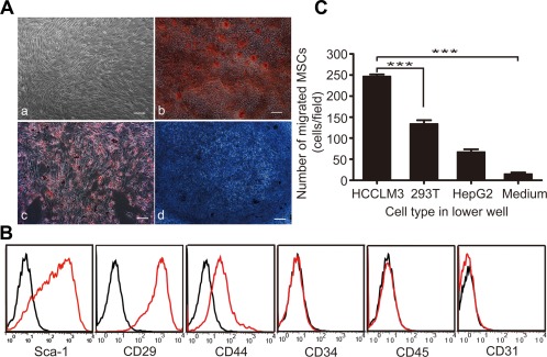
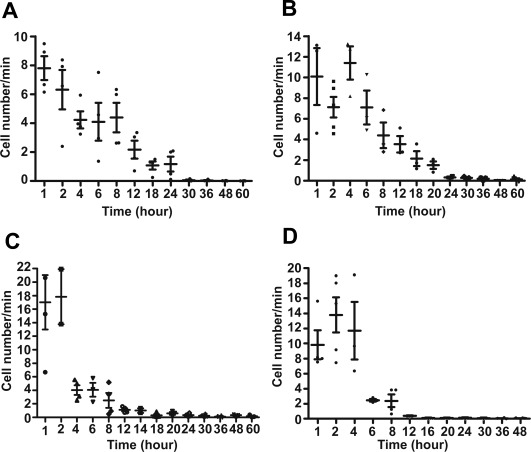
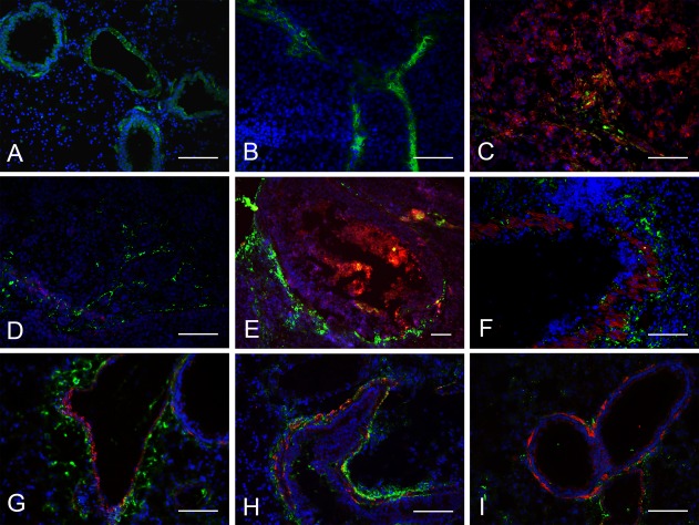
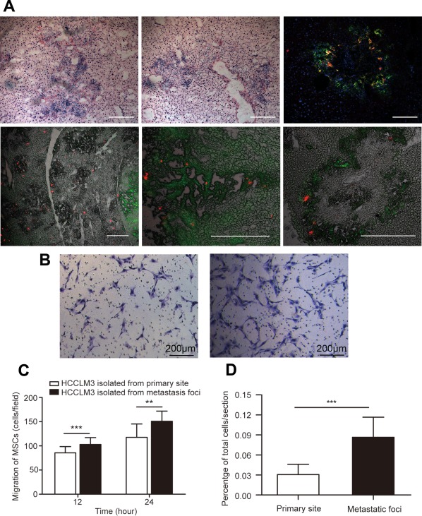
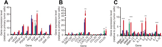
Bone marrow-derived mesenchymal stem cells (MSCs) can localize in injured, inflamed, and cancerous tissues after systemic infusion. However, the dynamic homing profile of MSCs in the peripheral blood is not well characterized. Here, using in vivo flow cytometry to noninvasively monitor the dynamics of fluorescence-labeled cells, we found different clearance kinetics of systemically infused MSCs between healthy and tumor mouse models. The circulation times of MSCs in healthy mice and mice with subcutaneous tumors, orthotopically transplanted liver tumors, or metastatic lung tumors were 30, 24, 18, and 12 hours, respectively, suggesting that MSCs actively home to tumor environments. MSCs infiltrated into hepatocellular carcinoma (HCC) sites and preferentially engrafted to micrometastatic regions both in vivo and in vitro. The expression of epidermal growth factor, CXCL9, CCL25, and matrix metalloproteinases-9 by HCC cells differed between primary tumor sites and metastatic regions. By characterizing the homing profiles of systemically perfused MSCs under physiological and cancerous conditions, these findings increase our understanding of the migration of MSCs from the circulation to tumor sites and constitute a basis for developing MSC-based anti-cancer therapeutic strategies. Stem Cells Translational Medicine 2017;6:1120-1131.

Keywords: Hepatocellular carcinoma; Homing; In vivo flow cytometry; Mesenchymal stem cells; Tumor xenograft model.


Figures

Figure 1
Figure 1
MSCs show typical characteristics and tropism to HCCLM3 cells in vitro. (Aa): Spindle‐shaped morphology of MSCs generated from adult mouse bone marrow. Differentiation capacity of MSCs into (Ab) osteoblasts (Alizarin Red S), (Ac) adipocytes (Oil Red O), and (Ad) chondrocytes (Toluidine Blue). Scale bar: 200 μm. (B): Transwell assay showed a greater migration of MSCs toward GFP‐HCCLM3 cells than toward HepG2 cells (control: 293T cells), ***, p < .001. (C): Cell surface markers of mouse MSCs. Histograms showing the expression of surface markers were plotted against controls. Abbreviations: MSCs, mesenchymal stem cells; Sca‐1, stem cell antigen; GFP, green fluorescent protein.
Figure 2
Figure 2
The circulation times of systemically infused mesenchymal stem cells as measured by in vivo flow cytometry were shorter in tumor mouse models than in healthy mice (n = 6 per group). The first measurement began within 5 minutes of i.v. infusion and lasted for 2 hours continuously. Later measurements were acquired at the same vessel at 2‐hour intervals until 8 hours and then 6‐hour intervals until 60 hours, with each measurement lasting 1 hour. The number of detected cells per minute is shown as a function of time following infusion in (A) healthy mice, (B) mice with subcutaneous tumors, (C) mice with orthotopic liver tumors, and (D) mice with metastatic lung tumors.
Figure 3
Figure 3
The depletion times of green fluorescent protein ‐mesenchymal stem cells (GFP‐MSCs) in the peripheral blood measured by conventional ex vivo flow cytometry were similar as those measured by in vivo flow cytometry. After MSC infusion into the tail vein, 150 μl blood samples were collected through the orbital venous plexus in (A) healthy mice, (B) mice with subcutaneous tumors, (C) mice with orthotopic liver tumors, and (D) mice with metastasized lung tumors at various time points. Data are shown as the proportion of GFP+ cells in 105 sampling cells (n = 6 mice per group).
Figure 4
Figure 4
Green fluorescent protein‐mesenchymal stem cells (GFP‐MSCs) migrated to the periphery of red fluorescent protein (RFP)‐HCCLM3 tumors and preferentially accumulated in micrometastatic regions as shown by immunofluorescence staining. In mice with subcutaneous tumors, MSCs became trapped in the (A) lungs, (B) liver, and (C) infiltrated across the subcutaneous tumor site. In mice with orthotopically transplanted tumors, MSCs engrafted to (D, E) the periphery of the primary tumor site and (F) metastatic regions. (HI) In mice with metastasized lung tumors, MSCs preferentially surrounded metastatic regions. Blue: 4′,6‐diamidino‐2‐phenylindole, green: MSCs, red: RFP‐HCCLM3 cells; scale bar: 100 μm.
Figure 5
Figure 5
MSCs preferentially infiltrated into micrometastatic regions both in vivo and in vitro. (A): Immunostaining revealed that MSCs homed to liver micrometastatic regions (upper panel). In 5(A), the lower panel shows different magnifications of DiD‐MSCs that migrated to intrahepatic metastatic foci. Scale bar: 100 μm. (B, C): When directly cocultured with HCCLM3 cells in transwell systems, MSCs showed greater migration toward GFP‐HCCLM3 cells extracted from micrometastatic regions (B, the right channel) than toward tumor cells extracted from the primary tumor site (B, the left channel). Scale bar: 200 μm. (D): The number of MSCs engrafted to the primary tumor site and metastatic regions was quantified from 15 random fields per section (three sections per mouse). Data are shown as the proportion of MSCs out of the total number of systemically infused MSCs per section. Original magnification, ×20. **, p < .05; ***, p < .001. Abbreviation: MSCs, mesenchymal stem cells.
Figure 6
Figure 6
The mRNA expression levels of three cytokines were higher in tumor cells extracted from metastatic regions than in cells extracted from primary tumor sites. mRNA expression of green fluorescent protein (GFP)‐HCCLM3 cells extracted from the primary orthotopic liver tumor site or metastatic regions and cultured alone or cocultured with mesenchymal stem cells (MSCs) in 0.4‐μm transwell systems for 24 hours. (A): Growth factors, (B) chemotactic factors, and (C) inflammatory factors. Black: HCCLM3 cells extracted from the primary tumor site, green: HCCLM3 cells extracted from micrometastatic regions, blue: HCCLM3 cells extracted from the primary tumor site and cocultured with MSCs, red: HCCLM3 cells extracted from micrometastatic regions and cocultured with MSCs. Data are shown as mean ± SEM; n = 3 per group; *, p < .05; **, p < .005; ***, p < .001. Abbreviations: bFGF, basic fibroblast growth factor; CCL, CC chemokine ligand; CXCL, CX chemokine ligand; EGF, epidermal growth factor; G‐CSF, granulocyte colony‐stimulating factor; GDF‐15, growth differentiation factor; GM‐CSF, granulocyte/macrophage colony‐stimulating factor; HGF, hepatocyte growth factor; IL, interleukin; MMP, matrix metalloproteinases; PDGF‐α, platelet‐derived growth factor; PDGF‐β, platelet‐derived growth factor‐β; TNF‐α, tumor necrosis factor; TGF‐β, transforming growth factor; VEGF, vascular endothelial growth factor.

Similar articles

Cited by

References

    1. Jemal A, Bray F, Center MM et al. Global cancer statistics. CA Cancer J Clin 2011;61:69–90. - PubMed
    1. Schäfer M, Werner S. Cancer as an overhealing wound: An old hypothesis revisited. Nat Rev Mol Cell Biol 2008;9:628–638. - PubMed
    1. Tlsty TD, Coussens LM. Tumor stroma and regulation of cancer development. Annu Rev Pathol Mech Dis 2006;1:119–150. - PubMed
    1. Quante M, Tu SP, Tomita H et al. Bone marrow‐derived myofibroblasts contribute to the mesenchymal stem cell niche and promote tumor growth. Cancer Cell 2011;19:257–272. - PMC - PubMed
    1. Erez N, Truitt M, Olson P et al. Cancer‐associated fibroblasts are activated in incipient neoplasia to orchestrate tumor‐promoting inflammation in an NF‐κB‐dependent manner. Cancer Cell 2010;17:135–147. - PubMed

Publication types