. 2017 Feb 16:8:14472.
doi: 10.1038/ncomms14472.

Tailored protein encapsulation into a DNA host using geometrically organized supramolecular interactions

Affiliations

Tailored protein encapsulation into a DNA host using geometrically organized supramolecular interactions

Andreas Sprengel et al. Nat Commun. .

Abstract

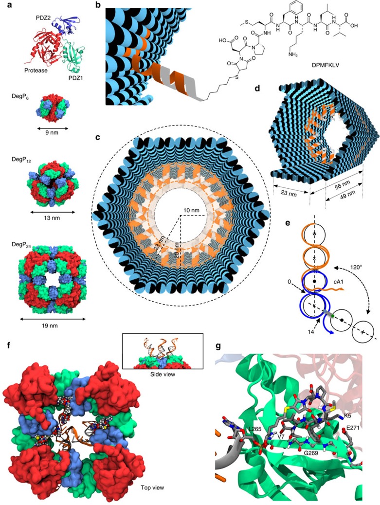
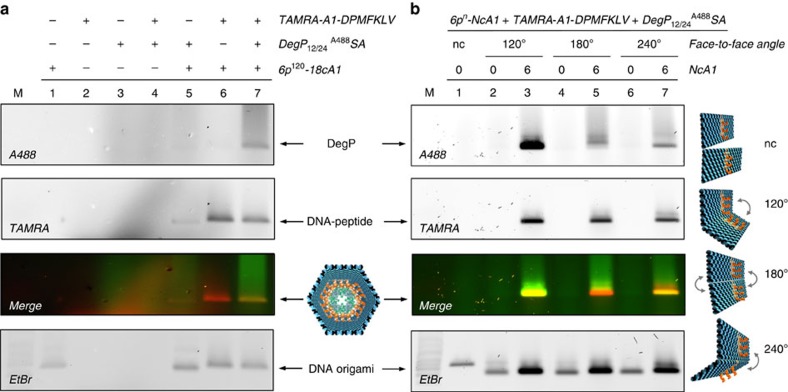
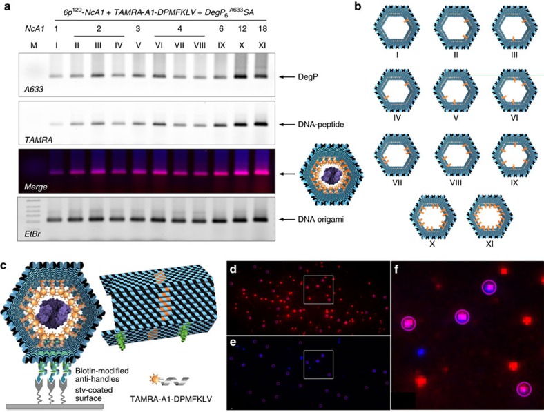
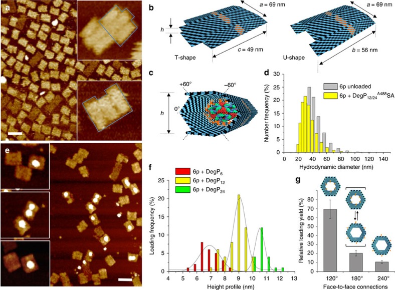
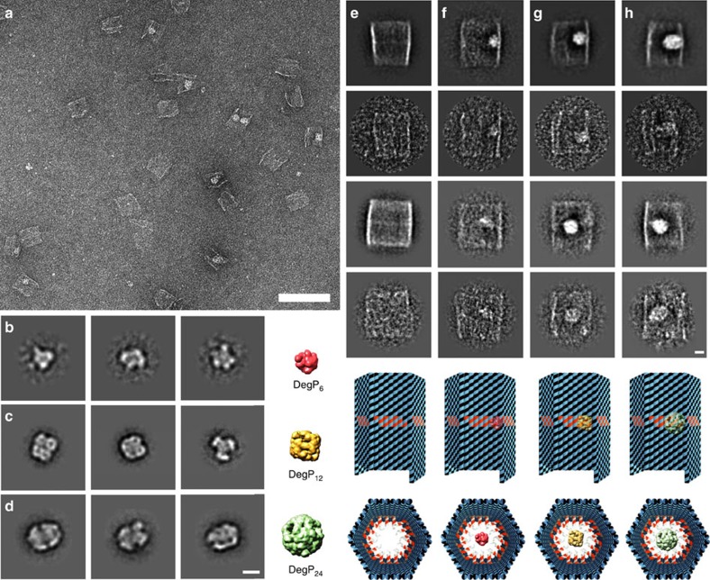
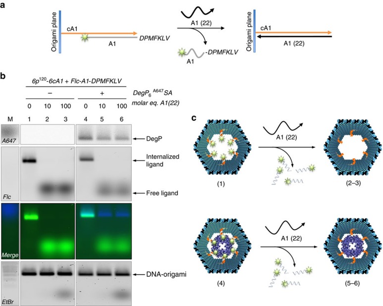
The self-organizational properties of DNA have been used to realize synthetic hosts for protein encapsulation. However, current strategies of DNA-protein conjugation still limit true emulation of natural host-guest systems, whose formation relies on non-covalent bonds between geometrically matching interfaces. Here we report one of the largest DNA-protein complexes of semisynthetic origin held in place exclusively by spatially defined supramolecular interactions. Our approach is based on the decoration of the inner surface of a DNA origami hollow structure with multiple ligands converging to their corresponding binding sites on the protein surface with programmable symmetry and range-of-action. Our results demonstrate specific host-guest recognition in a 1:1 stoichiometry and selectivity for the guest whose size guarantees sufficient molecular diffusion preserving short intermolecular distances. DNA nanocontainers can be thus rationally designed to trap single guest molecules in their native form, mimicking natural strategies of molecular recognition and anticipating a new method of protein caging.


Conflict of interest statement

The authors declare no competing financial interests.

Figures

Figure 1
Figure 1. The host–guest system and the encapsulation strategy.
(a) The DegP protein guest in its monomeric form is constituted by three domains: a protease (red), a PDZ1 (green) and a PDZ2 (blue) domain. The 6-, 12- and 24-mer DegP states have different symmetries and sizes (PDB codes are, respectively: 1KY9, 3OTP and 3CS0). (b) The DNA origami host is internally decorated with protruding DNA strands (orange helices) for further hybridization to complementary DNA–peptide conjugates (grey helices). (c) The host is made of six planar faces connected into a hexagonal prism with an edge and outer radius of 23 nm and a free inner room of ca. 10 nm in radius. (d) The host has two different lengths 49 and 56 nm, associated, respectively, to four short and two long opposite edges. (e) Schematic representation of the design strategy used to link adjacent faces at a fixed 120° angle, using out-of-plane crossovers. (f) Molecular dynamics simulations illustrate the binding of three DNA-DPMFKLV ligands to the PDZ1 domains of DegP24 (top view). The side view of a small region of the complex is shown in the panel. (g) Detailed view of the binding of the DNA-DPMFKLV ligand to the PDZ1 domain in the interior cavity of DegP24 (last frame of the MD simulations).
Figure 2
Figure 2. Gel electrophoresis analysis of DegP12/24A488 SA binding to the DNA host.
(a) Analysis of the single components of the complex (lanes 1–3, corresponding, respectively, to the DNA origami host, the TAMRA-labelled DNA–peptide conjugate and the DegP protein) as well as of their mutual interactions (lanes 4–7) indicates specific DNA–protein binding only in presence of the peptide (lane 7). (b) Binding of DegP12/24 to the 6p construct occurs only in presence of PAs (NcA1=6 in lanes 3, 5 and 7) and is dependent on their convergent (120°), randomly oriented (180°) or divergent (240°) arrangement. Only a convergent design of ligands (lane 3) leads to satisfactory yields of binding, whereas undefined (lane 5) or divergent (lane 7) ligand orientation is poorly efficient (yields are in a 8:1.4:1 ratio). As control, a DNA structure lacking the face-to-face connections (nc) has been used (lane 1). Lanes M contained 1 kbp DNA ladder (Roth). The DNA origami structures migrate between the 1.5 and 2.0 kbp bands of the ladder. Gel running conditions: 0.75% agarose in 1 × TBEMg buffer, 4 °C, 3 h at 80 V. Gel imaging was performed with a Typhoon FL900 upon illumination at selected wavelengths to allow detection of the protein (Alexa488), peptide ligand (TAMRA) and DNA (upon ethidium bromide staining). Full gels are shown in Supplementary Figs 34 and 43.
Figure 3
Figure 3. Binding of DegP6A633 SA to diverse 6p120 constructs.
(a) Agarose gel electrophoresis characterization of the binding of DegP6A633SA (labelled at genetically introduced cysteine residues with A633) to diverse 6p120 constructs, differing in the number (NcA1; from 1 to 18) and spatial arrangement of the ligands within the cavity (constructs I to XI, b). The results indicate successful binding for all constructs, with maximal efficiency for a radial distribution of ligands (constructs X and XI). Lane M contained 1 kbp DNA ladder (Roth). The DNA origami structures migrate between the 1.5 and 2.0 kbp bands of the ladder. Gel running conditions: 0.75% agarose in 1 × TBEMg buffer, 4 °C, 3 h at 80 V. Gel imaging was performed with a Typhoon FL900 upon illumination at selected wavelengths to allow detection of the protein (Alexa633), peptide ligand (TAMRA) and DNA (upon ethidium bromide staining). (c) Single-molecule fluorescence characterization of the 6p construct, bearing 18 convergent PAs hybridized with TAMRA-tagged peptide ligands and loaded with a DegP6A647SA protein. Molecules were immobilized on a coverslip surface and were measured using TIRF microscopy. TAMRA (red spots in d) and Alexa647 (blue spots in e) detection channels have been overlapped, indicating clear co-localization of the two species (violet spots showing energy transfer from the donor to the acceptor fluorophore in f, which shows a zoom-in view of the highlighted region in d,e).
Figure 4
Figure 4. AFM characterization of the unloaded and DegP-loaded host.
(a) The unloaded host deforms during AFM imaging in air, leading to a double-layered structure with one of two possible shapes: either a T or a U shape (b). Whereas the former results from the bending of the structure along the symmetry axis indicated as 0°, the latter is obtained from compression of the structure along the ±60° axes (c). Width and length of the obtained structures are as expected when compressing a correctly formed construct. (d) The hydrodynamic size distribution of the gel-purified DNA origami host either unloaded (grey bars) or loaded (yellow bars) with DegP12 protein was measured by dynamic light scattering, demonstrating correct formation of the hollow structure in solution. (e) Loading of the host with DegP protein mostly results in the appearance of single brighter dots in the centre of the structures, thus indicating successful protein binding in a 1:1 ratio. Scale bars, (a,e) 100 nm. (f) Analysis of the height profile of the structures revealed three distributions centred at ca. 7, 9 and 10.5 nm, corresponding, respectively, to encapsulation of the 6-, 12- and 24-mer (red, yellow and green bars, respectively). Preferential binding selectivity was observed for the 12-mer (more than 50% of the whole population). (g) Systematic analysis of the loading yield revealed most efficient protein encapsulation for a convergent design of multiple ligands (120°). Error bars indicate s.d.'s.
Figure 5
Figure 5. Negative stain EM of DegP and DegP-loaded DNA hosts.
(a) Representative digital micrograph area of negatively stained DegP12 and DegP24-loaded DNA cages. Scale bar, 100 nm. Representative class averages, each containing ∼50–100 particles, for DegP6 (b), DegP12 (c) and DegP24 (d). Scale bar, 10 nm. The simulated 3D models from the respective crystal structures downfiltered to a resolution of 15 Å are also indicated. PDB-ID codes for DegP6 (b), DegP12 (c) and DegP24 (d) are, respectively, 1ky9, 2zle and 3cS0. Two-dimensional analysis of empty (e), DegP6 (f), DegP12 (g) and DegP24 (h) loaded DNA origami hosts bearing 18 convergent PAs in their cavity (6p120-18cA1). Representative class averages, each containing ∼25–100 particles, and raw particle images of the corresponding classes are reported, together with a model of each construct, both in top and front view. Scale bar, 10 nm.
Figure 6
Figure 6. Electrophoretic analysis of single-strand displacement reactions.
(a) The A1-peptide ligands (grey strands) are only partially complementary to the cA1 strands (orange) protruding out of the origami plane, leaving six nucleobases available for attachment of a fully complementary sequence A1(22) (black), with consequent displacement of the A1-peptide conjugate. (b) Gel electrophoresis analysis of purified DNA cages, either unloaded (lanes 1–3) or DegP-loaded (lanes 4–6), upon treatment with 0, 10 or 100 equimolar amounts of A1(22). The reaction was let at 30 °C overnight. The results indicate protein-trapping despite successful displacement of the peptide ligands. Lane M contained 1 kbp DNA ladder (Roth). The DNA origami structures migrate between the 1.5 and 2.0 kbp bands of the ladder. Gel running conditions: 0.75% agarose in 1 × TBEMg, at 80 V for 2 h at 4 °C. The gel was scanned with a Typhoon FLA 9000 at different wavelengths to record the presence of DegP protein (Alexa647), peptide ligands (Flc) and DNA (upon ethidium bromide staining). Full gel is shown in Supplementary Fig. 58. (c) Schematic representation of the products obtained in each gel lane, before and after single-strand displacement reactions.

References

    1. Hurtley S. Spatial cell biology. Location, location, location. Introduction. Science 326, 1205 (2009). - PubMed
    1. Agapakis C. M., Boyle P. M. & Silver P. A. Natural strategies for the spatial optimization of metabolism in synthetic biology. Nat. Chem. Biol. 8, 527–535 (2012). - PubMed
    1. Chen A. H. & Silver P. A. Designing biological compartmentalization. Trends Cell Biol. 22, 662–670 (2012). - PubMed
    1. Conrado R. J., Varner J. D. & DeLisa M. P. Engineering the spatial organization of metabolic enzymes: mimicking nature's synergy. Curr. Opin. Biotechnol. 19, 492–499 (2008). - PubMed
    1. Lee H., DeLoache W. C. & Dueber J. E. Spatial organization of enzymes for metabolic engineering. Metab. Eng. 14, 242–251 (2012). - PubMed

Publication types