Comparative Study
. 2017 Mar 22;9(3):309-322.
doi: 10.1039/c6mt00226a.

Comparative studies of oxaliplatin-based platinum(iv) complexes in different in vitro and in vivo tumor models

Affiliations
Comparative Study

Comparative studies of oxaliplatin-based platinum(iv) complexes in different in vitro and in vivo tumor models

Simone Göschl et al. Metallomics. .

Abstract

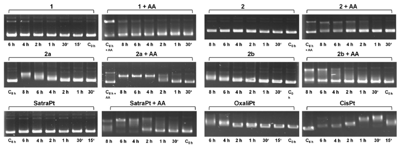
Using platinum(iv) prodrugs of clinically established platinum(ii) compounds is a strategy to overcome side effects and acquired resistances. We studied four oxaliplatin-derived platinum(iv) complexes with varying axial ligands in various in vitro and in vivo settings. The ability to interfere with DNA (pUC19) in the presence and absence of a reducing agent (ascorbic acid) was investigated in cell-free experiments. Cytotoxicity was compared under normoxic and hypoxic conditions in monolayer cultures and multicellular spheroids of colon carcinoma cell lines. Effects on the cell cycle were investigated by flow cytometry, and the capacity of inducing apoptosis was confirmed by flow cytometry and Western blotting. The anti-cancer activity of one complex was studied in vivo in immunodeficient and immunocompetent mice, and the platinum levels in various organs and the tumor after treatment were quantified. The results demonstrate that modification of the axial ligands can improve the cytotoxic potency. The complexes are able to interfere with plasmid DNA, which is enhanced by co-incubation with a reducing agent, and cause cell cycle perturbations. At higher concentrations, they induce apoptosis, but generate only low levels of reactive oxygen species. Two of the complexes increase the life span of leukemia (L1210) bearing mice, and one showed effects similar to oxaliplatin in a CT26 solid tumor model, despite the low platinum levels in the tumor. As in the case of oxaliplatin, activity in the latter model depends on an intact immune system. These findings show new perspectives for the development of platinum(iv) prodrugs of the anticancer agent oxaliplatin, combining bioreductive properties and immunogenic aspects.


Conflict of interest statement

Conflict of interests

The authors declare no conflict of interests.

Figures

Fig. 1
Fig. 1
Chemical structures of platinum(iv) complexes and reference compounds under investigation.
Fig. 2
Fig. 2
Electropherograms of dsDNA plasmid pUC19 incubated with 50 μM of platinum(iv) compounds 1, 2, 2a, 2b or satraplatin (in the absence or presence of 500 μM ascorbic acid, AA), oxaliplatin or cisplatin for different incubation times at 37 °C in TE buffer (pH = 7.8).
Fig. 3
Fig. 3
PI staining of the necrotic core (left) and immunohistochemical detection of HIF1α expression (right) in HCT15 and HCT116 spheroids with about 150 μm (A) and 400 μm (B) diameter (scale bars: 100 μm).
Fig. 4
Fig. 4
Cytotoxicity (concentration–effect curves) of complexes 2a, and 2b, oxaliplatin and satraplatin in HCT116 (white symbols) and HCT116oxR cells (black symbols) under different conditions, obtained by the fluorimetric resazurin assay after continuous exposure for 96 h.
Fig. 5
Fig. 5
Apoptosis induction by the investigated complexes after 24 h (upper row) and 48 h (lower row) of continuous exposure to either 2000 μM of 1, 2, 2a, 2b or 50 μM of oxaliplatin and satraplatin in HCT15, HCT116 and HCT116oxR cells. Cells were stained with annexin V–FITC/PI and quantified by flow cytometry. Percentages of viable (AV−/PI−), early apoptotic (AV+/PI−), late apoptotic (AV+/PI+) and necrotic (AV−/PI+) cells are means of at least three independent experiments evaluated by FlowJo software.
Fig. 6
Fig. 6
Immunoblots of caspase-induced cleavage of PARP in HCT15, HCT116 and HCT116oxR cells after continuous exposure for (A) 24 h with 2000 μM of 1, 2, 2a, 2b, 50 μM satraplatin and oxaliplatin; (B) 48 h with 1000 μM of 1, 2, 2a, 2b, 10 μM satraplatin and 50 μM oxaliplatin; and (C) 48 h with 2000 μM of 1, 2, 2a, 2b, 50 μM satraplatin and oxaliplatin. Representative blots from at least two independent experiments are depicted.
Fig. 7
Fig. 7
Fluorescence intensities of DCF as a measure of ROS levels in HCT116 (black symbols) and HCT116oxR (white symbols) cells recorded every 10 min during incubation with platinum(iv) complexes (2000 μM) and satraplatin (250 μM) for 2.5 h at 37 °C.
Fig. 8
Fig. 8
Kaplan–Meier plots of pilot experiments in the L1210 leukemia model. (A) Experiments on 2a were performed at the Cancer Research Institute, Slovak Academy of Sciences, Bratislava. The test compound was dissolved in DMSO and administered intraperitoneally at 20 mg kg−1 in a split-dose regimen on days 1, 2, and 3 after cell implantation (n = 2 per group). Control animals received DMSO i.p. (B) Experiments on 2b were performed at the Institute of Cancer Research, Medical University of Vienna, Austria. The test compound was dissolved in water and administered intraperitoneally at 20 mg kg−1 on days 1, 5, and 9 after cell implantation (n = 3 per group). Control animals received PBS i.p.
Fig. 9
Fig. 9
In vivo activity of 2b in male (A) BALB/c SCID (data shown are mean plus SEM; number of animals, 4 per group) and (B) BALB/c mice with subcutaneous CT26 tumors. Animals were treated with 20 mg kg−1 i.p. of 2b on days 4, 7, 11, and 14 (indicated by black arrows).
Fig. 10
Fig. 10
Average platinum concentrations (from treatment with 2b) in mouse tissues collected at day 15, quantified by ICP-MS.

References

    1. Reedijk J. Increased understanding of platinum anticancer chemistry. Pure Appl Chem. 2011;83:1709–1719.
    1. Wheate NJ, Walker S, Craig GE, Oun R. The status of platinum anticancer drugs in the clinic and in clinical trials. Dalton Trans. 2010;39:8113–8127. - PubMed
    1. Kelland L. The resurgence of platinum-based cancer chemotherapy. Nat Rev Cancer. 2007;7:573–584. - PubMed
    1. Jung Y, Lippard SJ. Direct cellular responses to platinum-induced DNA damage. Chem Rev. 2007;107:1387–1407. - PubMed
    1. Mehmood RK. Review of cisplatin and oxaliplatin in current immunogenic and monoclonal antibody treatments. Oncol Rev. 2014;8:256. - PMC - PubMed

Publication types

MeSH terms