Review
. 2017 Mar 14;17(6):1009-1023.
doi: 10.1039/c6lc01367h.

Bridging the gap: microfluidic devices for short and long distance cell-cell communication

Affiliations
Review

Bridging the gap: microfluidic devices for short and long distance cell-cell communication

Timothy Quang Vu et al. Lab Chip. .

Abstract

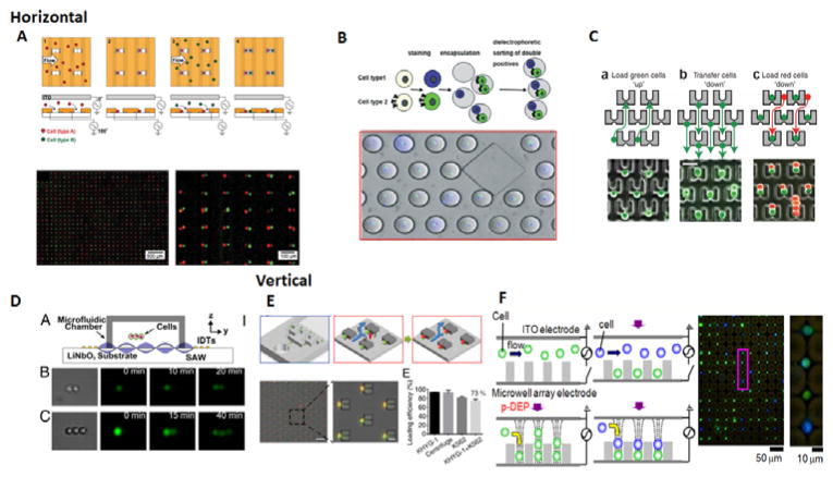
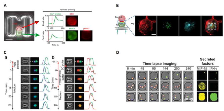
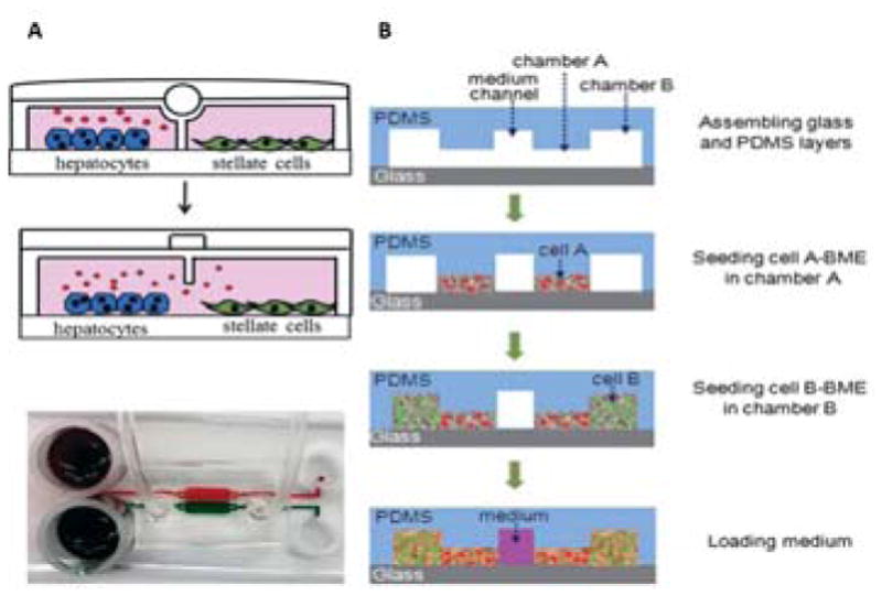
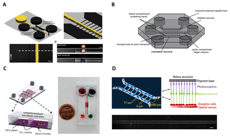
Cell-cell communication is a crucial component of many biological functions. For example, understanding how immune cells and cancer cells interact, both at the immunological synapse and through cytokine secretion, can help us understand and improve cancer immunotherapy. The study of how cells communicate and form synaptic connections is important in neuroscience, ophthalmology, and cancer research. But in order to increase our understanding of these cellular phenomena, better tools need to be developed that allow us to study cell-cell communication in a highly controlled manner. Some technical requirements for better communication studies include manipulating cells spatiotemporally, high resolution imaging, and integrating sensors. Microfluidics is a powerful platform that has the ability to address these requirements and other current limitations. In this review, we describe some new advances in microfluidic technologies that have provided researchers with novel methods to study intercellular communication. The advantages of microfluidics have allowed for new capabilities in both single cell-cell communication and population-based communication. This review highlights microfluidic communication devices categorized as "short distance", or primarily at the single cell level, and "long distance", which mostly encompasses population level studies. Future directions and translation/commercialization will also be discussed.


Figures

Figure 1
Figure 1
Horizontal and vertical cell pairing methods. A) Electrode based method of creating horizontal pairs. Positive dielectrophoresis(DEP) is used to trap each cell type(red and green) into the well wall, and negative DEP is used to manipulate the distances between cell pairs. B) Droplet-based approach for encapsulating two cells. Cell types 1 and 2 are stained and encapsulated, and the correct double positives can then be sorted out through DEP. C) Trap or structure based approached for horizontal cell pairing. The first cell(green) is captured upward into the gap and then transferred down into the trap. The second cell(red) is then flowed down to occupy the remaining trap space to be paired horizontally. D) Surface acoustic waves allow for control and manipulation of cell positions. A microfluidic chamber on top of the acoustic substrate with interdigital transducers allows for surface acoustic waves to travel into the device and connect the two or three cells together. E) Vertical cell pairing using DEP. Cells fall into the well using DEP and excess cells are removed away with a sucrose wash. Then the second cell is introduced and trapped using DEP on top of the first cell. F) Structure approach for vertical cell pairing. Cells are captured at the trap and fall into the micropit, which clears the trap. This allows for a second cell to be captured at the trap on top of the first cell. 1A. Reproduced from Ref with permission from the Royal Society of Chemistry. 1B. Reproduced from Ref with permission from the Royal Society of Chemistry. 1C. Reproduced from Ref with permission from Nature Publishing Group. 1D. Reproduced from Ref from the National Academy of Sciences. 1E. Reprinted with permission from Ref Copyright 2014 American Chemical Society 1F. Reproduced from Ref from The American Association of Immunologist.
Figure 2
Figure 2
Applications of cell pairing for communication studies. A) Immune cell heterogeneity studies. Single cell studies conducted on Ca2+ flux and activation between T cells(red) and B cells(green), where 225 individual cell pairs were tracked and analyzed based on CD8 expression(T cells) and MHCII and pMHC1 expression(B cells). The Ca2+ expression is measured by fluorescent signal over time. B) Imaging of the immunological synapse between a natural killer cell and a target cell. The authors were able to image the changes in localization between actin(red), perforin(green) and tubulin(blue) during the killing process. C) Cell fusion of cells using both chemical and electrical techniques. (a)Chemical fusion of mouse embryonic stem cells(green) to mouse embryonic(blue). (b)Electrical fusion of two 3T3s cells stained with DsRed and eGFP. Fusion is measured by the distance between fluorescence. D) Time lapse imaging and secretion analysis of NK cells interacting with target cells. NK cells(blue) can contact target cells(red) and subsequently kill the target cells(dead cells turn green through Sytox Green) Secreted factors are quantified by microengraving, where antibodies for MIP-1β and IFN-γ are bound to the bottom of the microwell. Secreted proteins are captured by the antibodies, and primary and fluorescent secondary antibodies for MIP-1β(yellow) and IFN-γ(green) bind. Concentration can be quantified by fluorescence intensity. 2A. Reproduced from Ref with permission from Nature Publishing Group. 2B. Reproduced from Ref from The American Association of Immunologist. 2C. Reproduced from Ref with permission from Nature Publishing Group. 2D. Reproduced from Ref with permission from the Royal Society of Chemistry.
Figure 3
Figure 3
Long distance 2D(A) or 3D(B) co-culture devices for secretion based communication. A) Cells are divided by a valve that can separate the two cells and control the secretion of cytokines(A, top). To make the cells communicate, the valve is opened so that secreted proteins and conditioned media can be shared between the populations(A, middle). This device could then be coupled with electrode sensors to monitor cell behavior. A schematic of the chip is shown to demonstrate compartments don’t mix when the valve is turned on(A, bottom) B) 3D cell co-culture using hydrogel culture. Cells are loaded in basement membrane extract(BME) gel to compartmentalize the cell. First, cell A(red) is introduced, then cell B(green) is introduced adjacent to cell A, but without mixing. The middle channel is filled with media to feed the cells. Cytokines, growth factors, and other secreted proteins can then pass through from one cell population to the next by diffusing through the gel. This also allows for cells to migrate through from one gel to the next. 3A. Reproduced from Ref with permission from the Royal Society of Chemistry. 3B. Reproduced from Ref with permission from the Royal Society of Chemistry.
Figure 4
Figure 4
Applications of neuron and ocular synapses studies A) Schematic of the microfluidic chip used for the visualizations and manipulation of synapses, where flow direction is achieved with the use of negative pressure thus by preventing diffusion into the microgrooves. The use of Alexa Fluor 488 allows for the observation of the difference in profile between perfusion and the washout step. B) Experimental setup of the 3 compartment microfluidic device used for synaptic competition. The use of microgrooves on the side chambers and ‘target’ neurons in the centre establishes the synaptic competition between axons to reach the central chamber. C) Schematics and actual image of neuron-astrocytes interaction microfluidic chip using food dyes to show the chambers and channels. This platform used for the first time simultaneously a combination of PEG, (polyethylene glycol), compartmentalized chambers and gen etically encoded calcium indicators for the analyses of neuron-neuron and neuron-astrocytes interaction. D) Microfluidic device schematics for retinal synapse regeneration above the retinal structure mimicked with the use of two chambers connected by 108 microchannels. Scale bar, 200μm. 4A. Reproduced from Ref with permission from Elsevier. 4B. Reproduced from Ref with permission from Elsevier. 4C. Reproduced from Ref with permission from Nature Publishing Group. 4D. Reproduced from Ref under a CC-BY license through Nature Publishing Group.
Figure 5
Figure 5
Applications of cell-to-cell protrusions in intercellular communication A) Image of the BloC-printing device displayed with ruler for scale and colored die for the visualization the channels B) Schematics of chip design with the symmetrical channel networks C) Flexibility of cell trapping with ribbon pattern as an example and body to body cell pairing with red and green cells with enlarged micrograph of cell pairs within dotted line D) Calcein transfer via GJIC in cell pairs with protrusion-to-protrusion and body-to-body pairing methods E) Forward and backwards protrusions generated by the fibroblasts after three hours of trapping. Fig 5. Reproduced from Ref with permission from the National Academy of Sciences.
Figure 6
Figure 6
Applications of bacterial cell communications A) Microfluidic device with a ratchet structure used bacterial cell-cell communication between two distinct motile bacterial cell types, the communication between cells was established via bridge-channels that connected both concentrator structures. The use of a ratchet structure prevents cells from moving through the bridge channels and insures only cell signaling molecules are able to cross the bridge. Both GFP and RFP cells were loaded at the same time and saw their concentration increased gradually over time B) Schematics of near cell-cell signaling with enhancement or attenuation of the signal molecules from the transmitter to reporter via modulating cells. The use of a 10cm flexible PTFE tube connecting the upstream transmitter cells with the reporter and enhancer/reducer cells downstream was essential for the detection gap time to mimic in vitro human digestion modules. The effluent solutions were collected either immediately after the transmitter cells upstream or allowed to flow downstream were they would encounter the remaining cell populations: test strain, reporter CT104, enhancer LW5, or reducer LW8. 6A. Reproduced from Ref with permission from the Royal Society of Chemistry. 6B. Reproduced from Ref with permission from the Royal Society of Chemistry.

References

    1. Mittelbrunn M, Sánchez-Madrid F. Nat Rev Mol Cell Biol. 2012;13:328–335. - PMC - PubMed
    1. Brücher BLDM, Jamall IS. Cell Physiol Biochem. 2014;34:213–243. - PubMed
    1. Hodgkin PD, Rush J, Gett AV, Bartell G, Hasbold J. Immunol Cell Biol. 1998;76:448–453. - PubMed
    1. Lai EC. Development. 2004;131:965 LP-973. - PubMed
    1. Malhotra A, Shanker A. Immunotherapy. 2011;3:1143–1166. - PMC - PubMed

Publication types