

Comment
doi: 10.7554/eLife.25001.
Micromanaging checkpoint proteins
Affiliations
- PMID: 28206949
- PMCID: PMC5313057
- DOI: 10.7554/eLife.25001
Item in Clipboard
Comment
Micromanaging checkpoint proteins
Elife.
.
Abstract
The kinase Mps1, long known to be the 'boss' in mitotic checkpoint signaling, phosphorylates multiple proteins in the checkpoint signaling cascade.
Keywords: E. coli; S. cerevisiae; S. pombe; biochemistry; cell biology; cell cycle; human; kinetochore; protein kinase; spindle checkpoint; xenopus.
Conflict of interest statement
The authors declare that no competing interests exist.
Figures
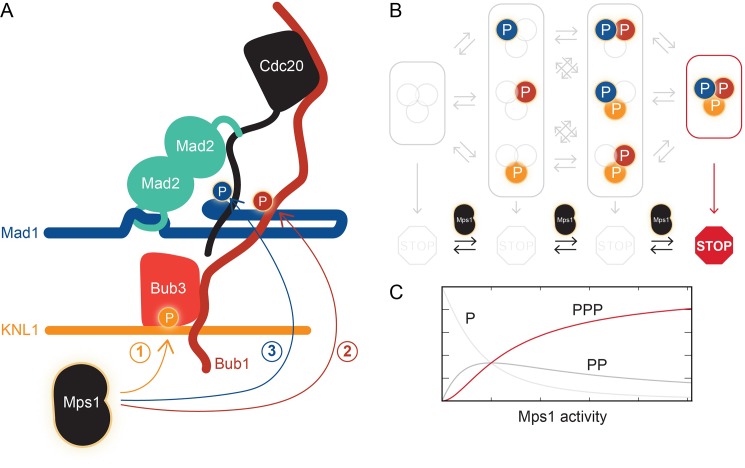
(A) Mps1 phosphorylates (P) three different proteins to promote the assembly of the mitotic checkpoint complex. It phosphorylates the kinetochore protein KNL1 to recruit the checkpoint protein complex Bub1-Bub3 to KNL1 (1). It phosphorylates Bub1, which allows this protein to interact with another checkpoint protein, Mad1 (2). It also phosphorylates Mad1, which promotes the binding of Mad2 to the regulatory protein Cdc20 (3). Ji et al. propose that phosphorylated Mad1 binds to Cdc20, thereby positioning the latter for capture by Mad2. (B) The checkpoint (represented by the STOP sign) is only active when Mps1 has phosphorylated all three proteins, KNL1, Bub1, and Mad1. (C) Checkpoint activity (y-axis) plotted as a function of Mps1 kinase activity (x-axis) for the phosphorylation of one (P), two (PP) or all three sites (PPP).
Comment on
-
A sequential multi-target Mps1 phosphorylation cascade promotes spindle checkpoint signaling.Elife. 2017 Jan 10;6:e22513. doi: 10.7554/eLife.22513. Elife. 2017. PMID: 28072388 Free PMC article.
References
Publication types
MeSH terms
Substances
LinkOut - more resources
Full Text Sources
Other Literature Sources
