Review
. 2017 Mar-Apr;23(2):118-126.
doi: 10.5152/dir.2016.16187.

Missed lung cancer: when, where, and why?

Affiliations
Review

Missed lung cancer: when, where, and why?

Annemilia Del Ciello et al. Diagn Interv Radiol. 2017 Mar-Apr.

Abstract

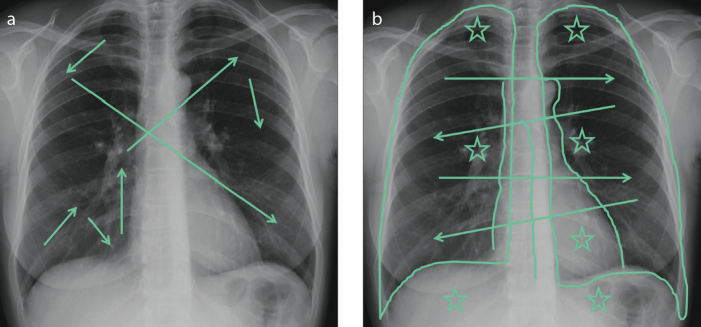
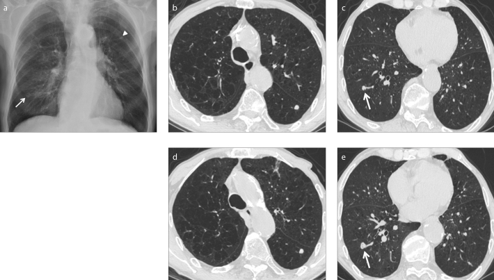
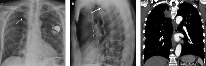
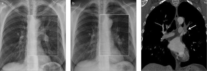
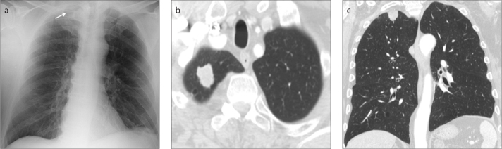
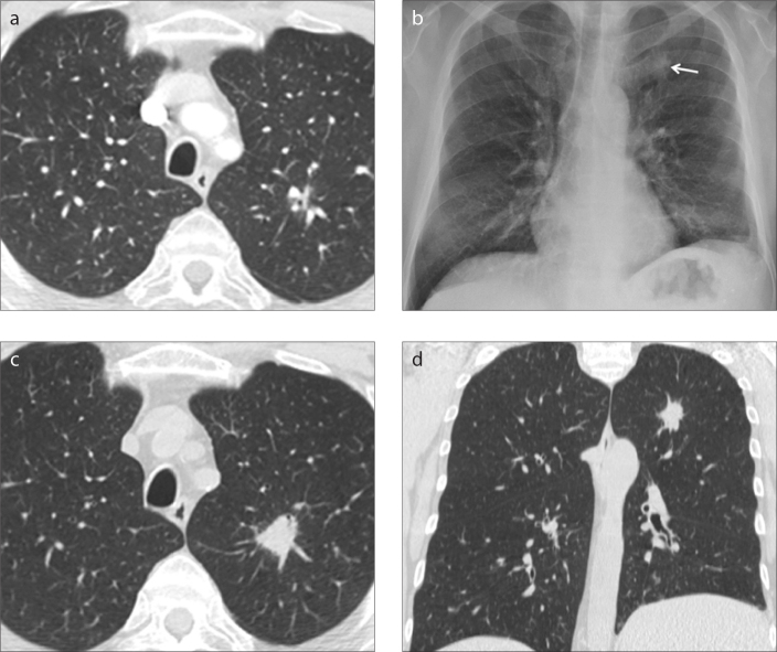
Missed lung cancer is a source of concern among radiologists and an important medicolegal challenge. In 90% of the cases, errors in diagnosis of lung cancer occur on chest radiographs. It may be challenging for radiologists to distinguish a lung lesion from bones, pulmonary vessels, mediastinal structures, and other complex anatomical structures on chest radiographs. Nevertheless, lung cancer can also be overlooked on computed tomography (CT) scans, regardless of the context, either if a clinical or radiologic suspect exists or for other reasons. Awareness of the possible causes of overlooking a pulmonary lesion can give radiologists a chance to reduce the occurrence of this eventuality. Various factors contribute to a misdiagnosis of lung cancer on chest radiographs and on CT, often very similar in nature to each other. Observer error is the most significant one and comprises scanning error, recognition error, decision-making error, and satisfaction of search. Tumor characteristics such as lesion size, conspicuity, and location are also crucial in this context. Even technical aspects can contribute to the probability of skipping lung cancer, including image quality and patient positioning and movement. Albeit it is hard to remove missed lung cancer completely, strategies to reduce observer error and methods to improve technique and automated detection may be valuable in reducing its likelihood.


Conflict of interest statement

Conflict of interest disclosure

The authors declared no conflicts of interest.

Figures

Figure 1. a, b
Figure 1. a, b
Frontal view chest X-rays showing examples of different scanning pathways. A messy scanning pattern (a) in which the reader’s gaze jumps to different areas of the lungs without any method (arrows). An ordered scan path (b), which covers all the lung zones symmetrically (arrows). The “blind zones” (apices, hila, retro-cardiac and sub-diaphragmatic spaces) (stars) and the mediastinal lines and stripes (lines) should always be checked carefully.
Figure 2. a–e
Figure 2. a–e
Missed lung cancer in an 85-year-old patient with severe emphysema. Routine postero-anterior (PA) chest X-ray (a) shows a nodular opacity projecting upon the posterior arch of the left sixth rib (arrowhead). Another nodular opacity in the right basal region (arrow) was not reported, probably because interpreted as a nipple (decision-making error) or because of satisfaction of search. CT exam performed immediately after the chest radiograph (b, c), confirms the presence of a nodule with regular margins in the apical segment of the left lower lobe (b). A nodule with lobulated margins is also visible in the right lower lobe (c, arrow) corresponding to the nodular opacity missed on chest radiograph. CT exam performed six months later (d, e) for nodule follow-up, shows the growth of the right lower lobe nodule (arrow), suggesting malignancy, later confirmed at biopsy. The diameter of the nodule in the left lower lobe is unchanged.
Figure 3. a–c
Figure 3. a–c
Missed lung cancer in a 75-year-old man who underwent chest X-ray after pacemaker positioning. A left iatrogenic pneumothorax was reported (a). An oval opacity in the posterior segment of the right upper lobe (arrows) was not identified by the radiologist (a, b). In the PA projection (a) the opacity was missed because of the superimposition with the fifth posterior rib arch crossing the first costo-sternal joint. In the lateral projection (b), the opacity was hidden by the upper thoracic vertebral bodies. CT performed a few days later (c) to rule out a pulmonary embolism, clearly depicted a soft tissue mass in the right upper lobe adjacent to the posterior mediastinal pleura.
Figure 4. a–c
Figure 4. a–c
Missed lung cancer in a 54-year-old heavy smoker patient with mild cough. PA chest X-ray (a) shows a slight left hilar increase in dimension and opacity with respect to the right hilum (square box), which was interpreted as negative. Comparison with a chest radiograph performed one year before (b) was of paramount importance to detect the subtle anatomic change at a second review of the images (square box). Coronal chest CT (c) at the mediastinum window setting documented solid tissue (arrow) in the left hilum occluding the apico-dorsal and anterior segmental bronchi of the left upper lobe, with subsequent segmental atelectasis and lobar volume loss.
Figure 5. a–c
Figure 5. a–c
Missed lung cancer with apical location in a bedside chest X-ray performed in a 66-year-old patient with brain metastasis. Antero-posterior (AP) radiograph (a), performed in the supine position, was reported as negative by the radiologist. The faint opacity in the right lung apex (white arrow) was hidden by the overlapping dense structures, such as ribs and right clavicle. CT exam later performed for staging purpose shows on axial (b) and coronal (c) images, a 2 cm nodular solid lesion with irregular margins in the apical segment of the right upper lobe, diagnosed as primary lung cancer.
Figure 6. a–d
Figure 6. a–d
Missed lung cancer on CT in a 63-year-old patient with hepatic cirrhosis hospitalized for ascites and complaining of cough and dyspnea. CT axial image (a) at lung window setting shows a small and poorly conspicuous lesion close to sub-segmental vessels of the apico-dorsal segment of the left upper lobe, that was not identified by the radiologist at the time of the exam, probably for its characteristics and location. Routine PA chest X-ray (b), performed two years later, shows a nodular opacity with irregular margins in the left apical region (arrow), identified by the radiologist. Axial (c) and coronal (d) CT images confirmed the presence of a 2 cm solid and not calcified nodule, with spiculated margins, located in the apico-dorsal segment of the left upper lobe, increased in diameter with respect to the previous CT exam and diagnosed as lung cancer.

References

    1. World Health Organization. Cancer. Fact Sheet Number 297. [Accessed July 10, 2015]. Available at: www.who.int/mediacentre/factsheets/fs297/en/Last updated July 2015.
    1. Groome PA, Bolejack V, Crowley JJ, et al. The IASLC Lung Cancer Staging Project: validation of the proposals for revision of the T, N, and M descriptors and consequent stage groupings in the forthcoming (seventh) edition of the TNM classification of malignant tumours. J Thorac Oncol. 2007;2:694–705. https://doi.org/10.1097/JTO.0b013e31812d05d5. - DOI - PubMed
    1. Shah PK, Austin JH, White CS, et al. Missed non-small cell lung cancer: radiographic findings of potentially resectable lesions evident only in retrospect. Radiology. 2003;226:235–241. https://doi.org/10.1148/radiol.2261011924. - DOI - PubMed
    1. White CS, Salis AI, Meyer CA. Missed lung cancer on chest radiography and computed tomography: imaging and medicolegal issue. J Thorac Imaging. 1999;14:63–68. https://doi.org/10.1097/00005382-199901000-00006. - DOI - PubMed
    1. Turkington PM, Kennan N, Greenstone MA. Misinterpretation of the chest x ray as a factor in delayed diagnosis of lung cancer. Postgrad Med J. 2002;78:158–160. https://doi.org/10.1136/pmj.78.917.158. - DOI - PMC - PubMed