. 2017 Apr 11;8(15):24869-24881.
doi: 10.18632/oncotarget.15276.

An autocrine/paracrine circuit of growth differentiation factor (GDF) 15 has a role for maintenance of breast cancer stem-like cells

Affiliations

An autocrine/paracrine circuit of growth differentiation factor (GDF) 15 has a role for maintenance of breast cancer stem-like cells

Asako Sasahara et al. Oncotarget. .

Abstract

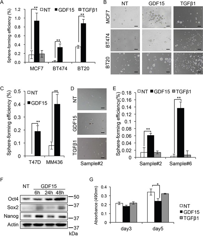
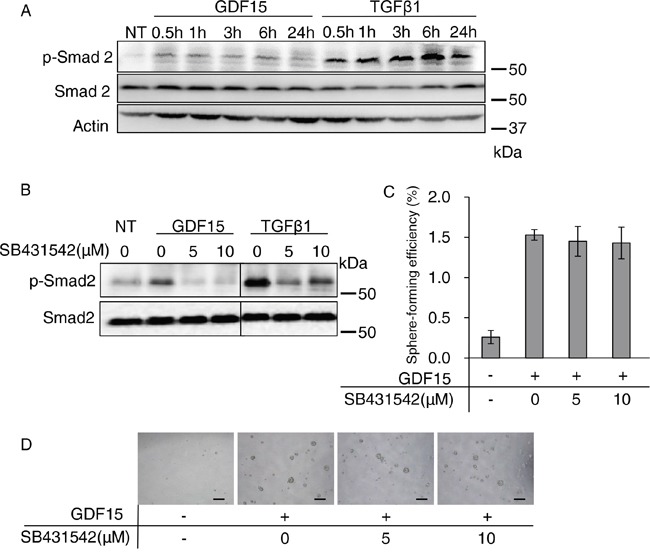
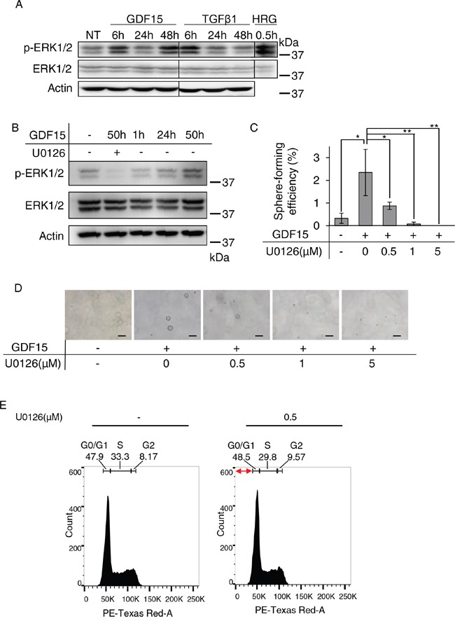
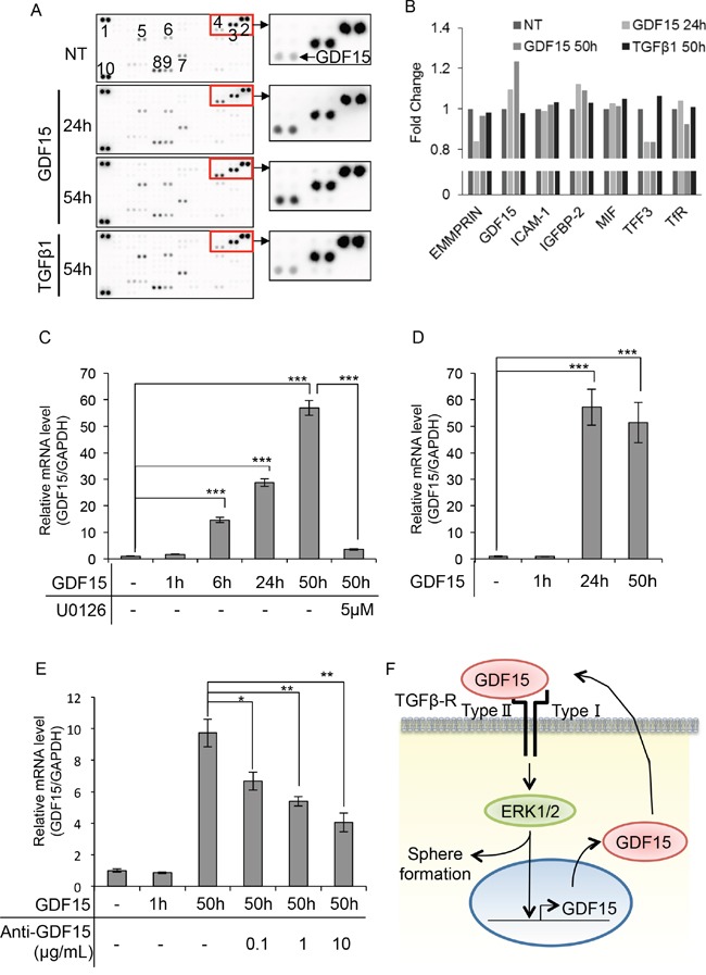
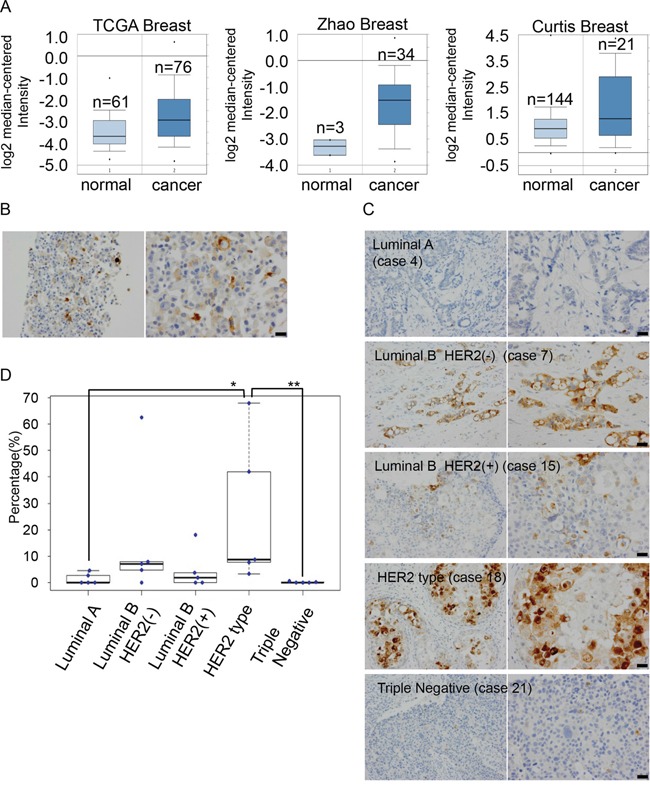
Cancer stem cells are thought to be responsible for tumor growth, recurrence, and resistance to conventional cancer therapy. However, it is still unclear how they are maintained in tumor tissues. Here, we show that the growth differentiation factor 15 (GDF15), a member of the TGFβ family, may maintain cancer stem-like cells in breast cancer tissues by inducing its own expression in an autocrine/paracrine manner. We found that GDF15, but not TGFβ, increased tumor sphere formation in several breast cancer cell lines and patient-derived primary breast cancer cells. As expected, TGFβ strongly stimulated the phosphorylation of Smad2. GDF15 also stimulated the phosphorylation of Smad2, but the GDF15-induced tumor sphere forming efficiency was not significantly affected by treatment with SB431542, an inhibitor of the TGFβ signaling. Although TGFβ transiently activated ERK1/2, GDF15 induced prolonged activation of ERK1/2. Treatment with U0126, an inhibitor of the MEK-ERK1/2 signaling, greatly inhibited the GDF15-induced tumor sphere formation. Moreover, cytokine array experiments revealed that GDF15, but not TGFβ, is able to induce its own expression; furthermore, it appears to form an autocrine/paracrine circuit to continuously produce GDF15. In addition, we found heterogeneous expression levels of GDF15 among cancer cells and in human breast cancer tissues using immunohistochemistry. This may reflect a heterogeneous cancer cell population, including cancer stem-like cells and other cancer cells. Our findings suggest that GDF15 induces tumor sphere formation through GDF15-ERK1/2-GDF15 circuits, leading to maintenance of GDF15high cancer stem-like cells. Targeting GDF15 to break these circuits should contribute to the eradication of tumors.

Keywords: ERK; TGFbeta; breast cancer; cancer stem cells; tumor spheres.


Conflict of interest statement

CONFLICTS OF INTEREST

No potential conflicts of interest were disclosed.

Figures

Figure 1
Figure 1. GDF15, but not TGFβ, efficiently induces tumor sphere formation in breast cancer cells
A. Tumor sphere assay of cell lines treated with GDF15 (200 ng/mL) or TGFβ1 (200 ng/mL). NT, not treated. n=4. **P < 0.01. B. Representative images of tumor spheres observed in (A). NT, not treated. Scale bar: 100 μm. C. Tumor sphere assay of cell lines treated with GDF15 (200 ng/mL). NT, not treated. n = 4, **P < 0.01. D. Representative images of tumor spheres derived from clinical sample #2, treated with GDF15 (200 ng/mL) or TGFβ1 (200 ng/mL). NT, not treated. Scale bar: 100 μm. E. The number of spheres in (D) and clinical sample #6 was counted and the percentage of sphere-forming efficiency was recorded. NT, not treated. n=4. **P < 0.01. F. Immunoblotting analysis of Oct4, Sox2, and Nanog expression in MCF7 cells treated with GDF15 (200 ng/mL). NT, not treated. Actin was used as a loading control. G. MTT assay using MCF7 cells. Cells were seeded to a 96-well plate and starved overnight prior to treatment with GDF15 (200 ng/mL) or TGFβ1 (200 ng/mL). The data was recorded at the indicated time. n=3. *P < 0.05.
Figure 2
Figure 2. Activation of the canonical Smad pathway is not required for GDF15-induced tumor sphere formation
A. Immunoblotting analysis of phosphorylated Smad2 (p-Smad2) and Smad2 expression in MCF7 cells treated with GDF15 (200 ng/mL) or TGFβ1 (200 ng/mL). NT, not treated. Actin was used as a loading control. B. Immunoblotting analysis of p-Smad2 and Smad2 expression in MCF7 cells. An indicated concentration of SB431542 was applied to the cells 60 minutes prior to treatment with GDF15 (200 ng/mL) or TGFβ1 (200 ng/mL). Cell lysates were collected 30 minutes after treatment with each ligand. NT, not treated. C. Tumor sphere assay of GDF15-treated (200 ng/mL) MCF7 cells in presence of the indicated concentrations of SB431542. n=4. D. Representative images of (C). Scale bar: 100 μm.
Figure 3
Figure 3. Sustained activation of ERK1/2 appears to be required for GDF15-induced tumor sphere formation
A. Immunoblotting analysis of phosphorylated ERK1/2 (p-ERK1/2) and ERK1/2 expression in MCF7 cells treated with GDF15 (200 ng/mL) or TGFβ1 (200 ng/mL). NT, not treated. Actin was used as a loading control. The lysate of MCF7 cells stimulated with heregulin (HRG) was used as a positive control. B. Immunoblotting analysis of p-ERK1/2 and ERK1/2 expression in MCF7 cells treated with GDF15 (200 ng/mL) in the presence or absence of U0126 (5 μM). C. Sphere formation assay of GDF15-treated (200 ng/mL) MCF7 cells in the presence or absence of the indicated concentration of U0126. n=4. **P < 0.01, *P < 0.05. D. Representative images of (C). Scale bar: 100 μm. E. Cell cycle analysis of MCF7 cells treated with GDF15 (200 ng/mL) in the presence or absence of U0126 (0.5 μM). Apoptotic cells are observed in the region indicated with the red arrows (the sub-G1 area).
Figure 4
Figure 4. GDF15 induces its own expression in a delayed time course
A. Cytokine array of MCF7 cells treated with GDF15 (200 ng/mL) or TGFβ1 (200 ng/mL). NT, not treated. The numbers of spots indicate cytokines: 1, 2, and 10, reference spots; 3, EMMPRIN; 4, GDF15; 5, ICAM-1; 6, IGFBP-2; 7, MIF; 8, TFF3; 9, TfR. B. The relative pixel densities of each spot detected in the cytokine array analyzed by ImageJ. The ratio of the number of treated cells to the number of un-treated cells for each treatment is shown in the graph. NT, not treated. n=2. C. Quantitative RT-PCR of MCF7 cells treated with GDF15 (200 ng/mL) in the presence or absence of U0126 (5 μM). Transcripts were collected at the indicated time. NT, not treated. n=3. ***P < 0.001. D. Quantitative RT-PCR of T47D cells treated with GDF15 (200 ng/mL). Transcripts were collected at the indicated time. NT, not treated. n=3. ***P < 0.001. E. Quantitative RT-PCR analysis of MCF7 cells treated with GDF15 (200 ng/mL) and the indicated concentration of the anti-GDF15 antibody. Transcripts were collected at the indicated time. NT, not treated. n=3. **P < 0.01. F. Estimated model of the GDF15-ERK1/2-GDF15 circuit in the promortion of tumor sphere formation.
Figure 5
Figure 5. Expression levels of GDF15 are heterogeneous among cancer cells in MCF7 cells and human breast cancer tissues
A. Analysis of the expression levels of GDF15 transcripts in Oncomine database. B. Immunohistochemical staining of GDF15 in MCF7 cells in paraffin blocks using an anti-GDF15 antibody. Left, original magnification 200x. Right, scale bar: 20 μm. C. Immunohistochemical staining of GDF15 in various subtypes of breast cancer tissues. Left, original magnification 400x. Right, scale bar: 20 μm. D. Box plots of GDF15 expression among 25 clinical breast cancer tissues that include 5 cases in each subtype. **P < 0.01, *P < 0.05.

References

    1. Torre LA, Bray F, Siegel RL, Ferlay J, Lortet-Tieulent J, Jemal A. Global cancer statistics, 2012. CA Cancer J Clin. 2015;65:87–108. - PubMed
    1. Goldhirsch A, Winer EP, Coates AS, Gelber RD, Piccart-Gebhart M, Thurlimann B, Senn HJ, Panel m. Personalizing the treatment of women with early breast cancer highlights of the St Gallen International Expert Consensus on the Primary Therapy of Early Breast Cancer 2013. Ann Oncol. 2013;24:2206–23. - PMC - PubMed
    1. Kreso A, Dick JE. Evolution of the cancer stem cell model. Cell Stem Cell. 2014;14:275–91. - PubMed
    1. Reya T, Morrison SJ, Clarke MF, Weissman IL. Stem cells, cancer, and cancer stem cells. Nature. 2001;414:105–11. - PubMed
    1. Badve S, Nakshatri H. Breast-cancer stem cells-beyond semantics. Lancet Oncol. 2012;13:e43–8. - PubMed

Substances