

Cross-Linked Micellar Spherical Nucleic Acids from Thermoresponsive Templates
- PMID: 28207251
- PMCID: PMC5493153
- DOI: 10.1021/jacs.6b13359
Cross-Linked Micellar Spherical Nucleic Acids from Thermoresponsive Templates
Abstract
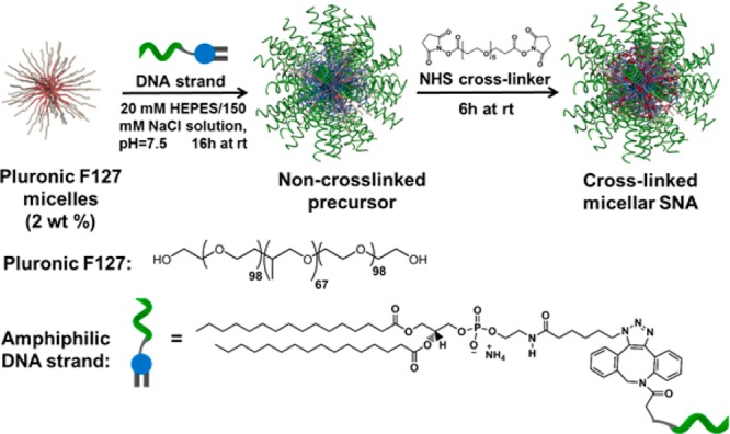
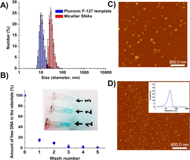
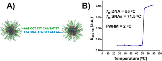
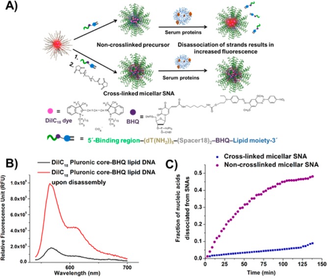
A one-pot synthesis of micellar spherical nucleic acid (SNA) nanostructures using Pluronic F127 as a thermoresponsive template is reported. These novel constructs are synthesized in a chemically straightforward process that involves intercalation of the lipid tails of DNA amphiphiles (CpG motifs for TLR-9 stimulation) into the hydrophobic regions of Pluronic F127 micelles, followed by chemical cross-linking and subsequent removal of non-cross-linked structures. The dense nucleic acid shell of the resulting cross-linked micellar SNA enhances their stability in physiological media and facilitates their rapid cellular internalization, making them effective TLR-9 immunomodulatory agents. These constructs underscore the potential of SNAs in regulating immune response and address the relative lack of stability of noncovalent constructs.
Conflict of interest statement
The authors declare no competing financial interest.
Figures
Similar articles
-
Structures and Applications of Nucleic Acid-Based Micelles for Cancer Therapy.Int J Mol Sci. 2023 Jan 13;24(2):1592. doi: 10.3390/ijms24021592. Int J Mol Sci. 2023. PMID: 36675110 Free PMC article. Review.
-
Cytotoxicity and internalization of Pluronic micelles stabilized by core cross-linking.J Control Release. 2014 Dec 28;196:87-95. doi: 10.1016/j.jconrel.2014.10.001. Epub 2014 Oct 13. J Control Release. 2014. PMID: 25307996
-
Preparation of novel ferrocene-based shell cross-linked thermoresponsive hybrid micelles with antitumor efficacy.J Phys Chem B. 2010 Apr 29;114(16):5309-14. doi: 10.1021/jp100901p. J Phys Chem B. 2010. PMID: 20369878
-
Photochemical Stabilization of Self-Assembled Spherical Nucleic Acids.Small. 2025 Feb;21(7):e2407742. doi: 10.1002/smll.202407742. Epub 2025 Jan 10. Small. 2025. PMID: 39790078 Free PMC article.
-
Analysis of protein-nucleic acid interactions by photochemical cross-linking and mass spectrometry.Mass Spectrom Rev. 2002 May-Jun;21(3):163-82. doi: 10.1002/mas.10024. Mass Spectrom Rev. 2002. PMID: 12476441 Review.
Cited by
-
Structures and Applications of Nucleic Acid-Based Micelles for Cancer Therapy.Int J Mol Sci. 2023 Jan 13;24(2):1592. doi: 10.3390/ijms24021592. Int J Mol Sci. 2023. PMID: 36675110 Free PMC article. Review.
-
Spherical nucleic acids: Organized nucleotide aggregates as versatile nanomedicine.Aggregate (Hoboken). 2022 Feb;3(1):e120. doi: 10.1002/agt2.120. Epub 2021 Sep 14. Aggregate (Hoboken). 2022. PMID: 35386748 Free PMC article.
-
DNA Nanostructure as an Efficient Drug Delivery Platform for Immunotherapy.Front Pharmacol. 2020 Jan 28;10:1585. doi: 10.3389/fphar.2019.01585. eCollection 2019. Front Pharmacol. 2020. PMID: 32063844 Free PMC article. Review.
-
Nanoarchitectonics of Spherical Nucleic Acids with Biodegradable Polymer Cores: Synthesis and Evaluation.Materials (Basel). 2022 Dec 13;15(24):8917. doi: 10.3390/ma15248917. Materials (Basel). 2022. PMID: 36556721 Free PMC article.
-
Spherical Nucleic Acids as Precision Therapeutics for the Treatment of Cancer-From Bench to Bedside.Cancers (Basel). 2022 Mar 23;14(7):1615. doi: 10.3390/cancers14071615. Cancers (Basel). 2022. PMID: 35406387 Free PMC article. Review.
References
Publication types
MeSH terms
Substances
Grants and funding
LinkOut - more resources
Full Text Sources
Other Literature Sources
Research Materials
