. 2017 Feb 16;13(2):e1006632.
doi: 10.1371/journal.pgen.1006632. eCollection 2017 Feb.

JMJD-5/KDM8 regulates H3K36me2 and is required for late steps of homologous recombination and genome integrity

Affiliations

JMJD-5/KDM8 regulates H3K36me2 and is required for late steps of homologous recombination and genome integrity

Pier Giorgio Amendola et al. PLoS Genet. .

Abstract

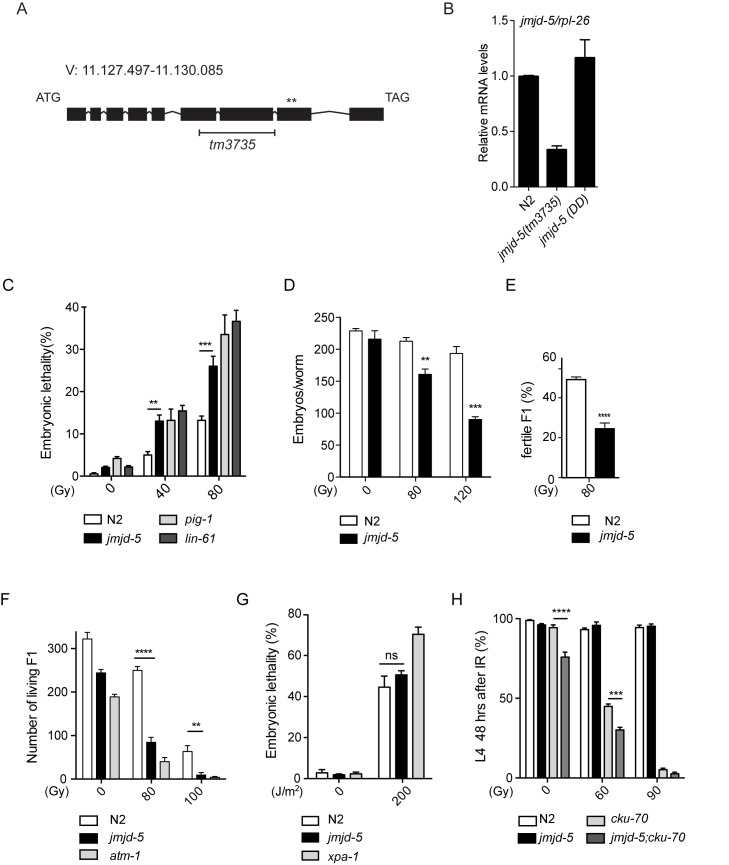
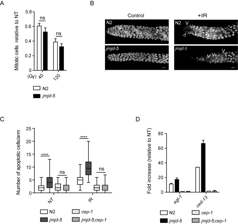
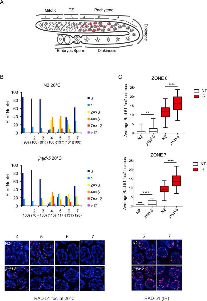
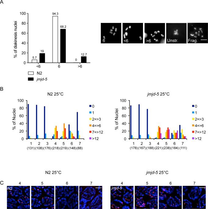
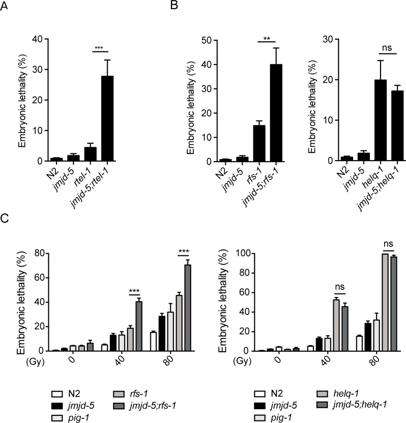
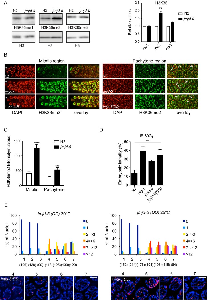
The eukaryotic genome is organized in a three-dimensional structure called chromatin, constituted by DNA and associated proteins, the majority of which are histones. Post-translational modifications of histone proteins greatly influence chromatin structure and regulate many DNA-based biological processes. Methylation of lysine 36 of histone 3 (H3K36) is a post-translational modification functionally relevant during early steps of DNA damage repair. Here, we show that the JMJD-5 regulates H3K36 di-methylation and it is required at late stages of double strand break repair mediated by homologous recombination. Loss of jmjd-5 results in hypersensitivity to ionizing radiation and in meiotic defects, and it is associated with aberrant retention of RAD-51 at sites of double strand breaks. Analyses of jmjd-5 genetic interactions with genes required for resolving recombination intermediates (rtel-1) or promoting the resolution of RAD-51 double stranded DNA filaments (rfs-1 and helq-1) suggest that jmjd-5 prevents the formation of stalled postsynaptic recombination intermediates and favors RAD-51 removal. As these phenotypes are all recapitulated by a catalytically inactive jmjd-5 mutant, we propose a novel role for H3K36me2 regulation during late steps of homologous recombination critical to preserve genome integrity.


Conflict of interest statement

The authors have declared that no competing interests exist.

Figures

Fig 1
Fig 1. jmjd-5 mutant animals are hypersensitive to IR.
(A) Genomic structure of the jmjd-5 gene. Dark boxes represent coding sequences and lines represent introns. The black H-shaped bar specifies the deletion in the tm3735 allele and the asterisks indicate the position of the two amino acids (H484 and D486) located in the JmjC domain, mutagenized in jmjd-5(DD). (B) Quantification of jmjd-5 mRNA levels by qRT-PCR in the indicated strains. rpl-26 is used as internal control. (C) Percent of embryonic lethality in the indicated strains observed after irradiation of young adult worms (24 h post L4) with 0, 40, or 80 Gy of IR. (D) Brood size in N2 and jmjd-5(tm3735) after IR treatment of young adults with 0, 80, or 120 Gy. Deposited embryos are counted from the time of irradiation. (E) Percentage of fertile animals in N2 and jmjd-5(tm3735) (evaluated by the presence of embryos in the uterus) among the living F1 progeny of irradiated mothers. (F) Number of living F1 animals in the indicated strains after irradiation of L1 larvae with 0, 80, or 100 Gy of IR. L1 were irradiated and the total number of living F1 offspring that passed the L1 stage is reported. (G) Percent of embryonic lethality in the indicated strains observed after treatment of young adult worms with 0 or 200 J/m2 of UV. (H) Percent of irradiated L1 of the indicated strains that developed to the L4 stage in 48 hours. In C-H, pig-1(gm344), lin-61(n3809), atm-1(gk186), cku-70(tm1524) and xpa-1(ok698) are used as positive controls. In B-H, data are represented as means ± sem from at least 3 biological independent experiments. ****p< 0.0001, ***p<0.005, **p<0.01, n.s. = not significant, with two-tailed, unpaired t-test.
Fig 2
Fig 2. jmjd-5 is not required for DNA damage checkpoints activation.
(A) Number of mitotic cells in N2 and jmjd-5(tm3735) after irradiation of young adults with 40 and 120 Gy, counted 24 h after IR, relative to the non treated (NT) animals. (B) Representative images of germlines dissected from N2 and jmjd-5 animals, 24 hours after irradiation with 0 (control) or 80 Gy (+IR), stained with DAPI. Arrowheads indicate arrested and enlarged nuclei. Scale bar: 5 μm. (C) Quantification of apoptotic cells in the indicated strains after staining with SYTO12, 24 hours after IR treatment (80Gy). NT, not treated. Between 40 and 78 animals were scored for genotype and condition. ****p< 0.0001, n.s. = not significant, with two-tailed, unpaired t-test. (D) Relative mRNA expression levels of the pre-apoptotic genes egl-1 and ced-13 in the indicated strains, 24 hours after IR treatment, measured by qPCR. Values are normalized to untreated samples and act-1 is used as internal control. Graph is an average of two biological independent experiments ± sem. cep-1(gk138) is used as control.
Fig 3
Fig 3. Aberrant RAD-51 staining in jmjd-5 mutant animals.
(A) Schematic representation of a C. elegans gonad arm, showing the position of the 7 zones scored for RAD-51 foci. TZ is the transition zone. Red dots represent RAD-51 foci distribution, according to literature. (B) Top. Histograms showing the quantification of RAD-51 foci in the extracted germlines of N2 and jmjd-5(tm3735) animals. Numbers in parenthesis indicate the number of nuclei analyzed. Bottom. Representative images of RAD-51 (red) and DAPI (blue) staining in germlines dissected from N2 and jmjd-5(tm3735) animals grown at 20°C. Nuclei from zones 4, 5, 6 and 7 are shown. (C) Top. Quantification of RAD-51 foci per nucleus in zones 6 and 7 of N2 and jmjd-5(tm3735) germlines, treated or not with IR (30Gy, 6 hours after the IR). At least 40 nuclei, from a minimum of 4 germlines for each genotype, were quantified for each zone and genotype. ****p< 0.0001, **p< 0.001, with two tailed unpaired t-test. Bottom. Representative images of RAD-51 (red) and DAPI (blue) staining in germlines dissected from N2 and jmjd-5 animals, 6h after IR (30Gy). Nuclei from zones 6 and 7 are shown. In (B) and (C), each image shows a projection of multiple z-stacks (0.2 μm spacing) of the entire nuclei. 100X magnification, scale bar 2 μm.
Fig 4
Fig 4. Role of jmjd-5 in meiotic homologous recombination.
(A) Left. Quantification of DAPI-stained bodies at diakinesis in N2 and jmjd-5 mutants, grown at 25°C for five generations. Nuclei were divided in three classes based on the number of DAPI-stained bodies/nucleus, presented in the x-axis. The y-axis represents the percentage of nuclei in each class. Numbers of nuclei scored were 122 for N2 and 110 for jmjd-5(tm3735). Right: representative images of DAPI-stained nuclei at diakinesis in N2 and jmjd-5(tm3735) mutants grown at 25°C for five generations. The number within each panel indicates the number of DAPI-stained bodies detected. Unstr: Unstructured chromosomes, Frag: Fragmented chromosomes. Scale bar 5 μm. (B) Histograms showing the quantification of RAD-51 foci in germlines extracted from N2 (left) and jmjd-5(tm3735) (right) animals grown at 25°C for five generations. Numbers in parenthesis indicate the number of nuclei analyzed. (C) Representative images of RAD-51 (red) and DAPI (blue) staining in dissected germlines of N2 (left) and jmjd-5(tm3735) (right) grown at 25°C for five generations. Nuclei from zones 4, 5, 6 and 7 are shown. In (A) and (C) each panel shows a projection of multiple z-stacks (0.2 μm spacing) of the entire nucleus. 100X magnification, scale bar 2 μm.
Fig 5
Fig 5. Genetic interaction analysis of jmjd-5 with rtel-1, helq-1 and rfs-1.
(A) Percentage of embryonic lethality measured in the indicated strains, grown at 25°C for one generation. Embryos from adult animals grown at 20°C were plated at 25°C and their ability, once adult, to produce viable progeny was measured. (B) Percentage of embryonic lethality measured in the indicated strains, grown at 25°C for one generation as described in (A). (C) Percentage of embryonic lethality in the indicated strains after treatment of young adults worms with 0, 40 and 80 Gy. pig-1(gm344) is used as positive control. Data are represented as means ± sem from at least 3 biological independent experiments. ***p<0.005, **p<0.01, n.s. = not significant, with two-tailed, unpaired t-test.
Fig 6
Fig 6. The catalytic activity of JMJD-5 is required for proper HR.
(A) Left. Western blots of the indicated histone modifications using lysates from N2 and jmjd-5(tm3735) animals. H3 is used as loading control. Right. Quantification of H3K36 methylation levels, normalized to H3. Data are presented as a mean ± sem of three independent biological experiments. **p<0.01 with two tailed unpaired t-test. (B) Representative images of indicated germline regions of N2, jmjd-5(tm3735) and jmjd-5(DD) animals stained with DAPI (red) and H3K36me2 antibody (green). Overlay is on the right. Asterisks indicate distal tip cells, arrowheads indicate regions devoid of H3K36me2. (C) Quantification of average H3K36me2 intensity per nucleus in mitotic and pachytene regions of N2 and jmjd-5 mutants. y-axis shows fluorescence intensity, expressed in arbitrary units. At least 10 gonads were quantified for each strain. Data are presented as mean ± sem. ****p< 0.0001, **p<0.01 with two tailed unpaired t-test. (D) Percent embryonic lethality in the indicated strains after treatment of young adults worms with 80 Gy. pig-1(gm344) is used as positive control. Data are represented as means ± sem of two independent biological experiments. **p<0.01 with two tailed unpaired t-test. (E) Top. Histograms showing the quantification of RAD-51 foci in germlines extracted from jmjd-5(DD) animals grown at 20°C (left) or at 25°C (right) for five generations. Numbers in parenthesis indicate the number of nuclei analyzed. Bottom. Representative images of RAD-51 (red) and DAPI (blue) staining in germlines of jmjd-5(DD) at 20°C (left) and 25°C (right). Nuclei from zones 4, 5, 6 and 7 are shown. In (B) and (E) each panel shows a projection of multiple z-stacks (0.2 μm spacing) of the entire nucleus. 100X magnification, scale bar 2 μm.

Similar articles

Cited by

References

    1. Ceccaldi R, Rondinelli B, D'Andrea AD. Repair Pathway Choices and Consequences at the Double-Strand Break. Trends Cell Biol. 2016;26(1):52–64. 10.1016/j.tcb.2015.07.009 - DOI - PMC - PubMed
    1. Jackson SP, Bartek J. The DNA-damage response in human biology and disease. Nature. 2009;461(7267):1071–8. 10.1038/nature08467 - DOI - PMC - PubMed
    1. Polo SE, Jackson SP. Dynamics of DNA damage response proteins at DNA breaks: a focus on protein modifications. Genes & development. 2011;25(5):409–33. - PMC - PubMed
    1. Heyer W-D, Ehmsen KT, Liu J. Regulation of homologous recombination in eukaryotes. Annual review of genetics. 2010;44:113 10.1146/annurev-genet-051710-150955 - DOI - PMC - PubMed
    1. Symington LS, Gautier J. Double-strand break end resection and repair pathway choice. Annual review of genetics. 2011;45:247–71. 10.1146/annurev-genet-110410-132435 - DOI - PubMed

Publication types

MeSH terms