. 2017 Feb 16;13(2):e1006586.
doi: 10.1371/journal.pgen.1006586. eCollection 2017 Feb.

Discovery of a new family of relaxases in Firmicutes bacteria

Affiliations

Discovery of a new family of relaxases in Firmicutes bacteria

Gayetri Ramachandran et al. PLoS Genet. .

Abstract

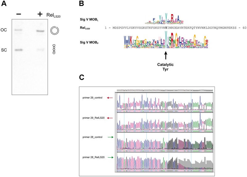
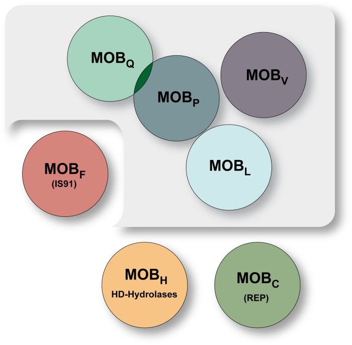
Antibiotic resistance is a serious global problem. Antibiotic resistance genes (ARG), which are widespread in environmental bacteria, can be transferred to pathogenic bacteria via horizontal gene transfer (HGT). Gut microbiomes are especially apt for the emergence and dissemination of ARG. Conjugation is the HGT route that is predominantly responsible for the spread of ARG. Little is known about conjugative elements of Gram-positive bacteria, including those of the phylum Firmicutes, which are abundantly present in gut microbiomes. A critical step in the conjugation process is the relaxase-mediated site- and strand-specific nick in the oriT region of the conjugative element. This generates a single-stranded DNA molecule that is transferred from the donor to the recipient cell via a connecting channel. Here we identified and characterized the relaxosome components oriT and the relaxase of the conjugative plasmid pLS20 of the Firmicute Bacillus subtilis. We show that the relaxase gene, named relLS20, is essential for conjugation, that it can function in trans and provide evidence that Tyr26 constitutes the active site residue. In vivo and in vitro analyses revealed that the oriT is located far upstream of the relaxase gene and that the nick site within oriT is located on the template strand of the conjugation genes. Surprisingly, the RelLS20 shows very limited similarity to known relaxases. However, more than 800 genes to which no function had been attributed so far are predicted to encode proteins showing significant similarity to RelLS20. Interestingly, these putative relaxases are encoded almost exclusively in Firmicutes bacteria. Thus, RelLS20 constitutes the prototype of a new family of relaxases. The identification of this novel relaxase family will have an important impact in different aspects of future research in the field of HGT in Gram-positive bacteria in general, and specifically in the phylum of Firmicutes, and in gut microbiome research.


Conflict of interest statement

The authors have declared that no competing interests exist.

Figures

Fig 1
Fig 1. A 17 residue region of the deduced protein sequence of pLS20cat gene 58 shows similarity to the motif III regions of MOBP, MOBQ and MOBV type relaxases.
pLS20cat has been sequenced independently in our lab and in the lab of M. Itaya (Keio University, Japan) who deposited the sequence in public databases where it was given the accession number NC_015148.1. pLS20cat protein p58, according to our nomenclature, has been assigned the accession number YP_004243525.1. Residues 141–171 of the deduced pLS20cat protein p58 are aligned with the consensus sequences, represented as Weblogos, of the motif III region of relaxases belonging to the MOBP, MOBQ and MOBV family. The consensus sequence of the motif III regions of the published relaxases of each MOB family [24] was identified here with the motif-identification program MEME [27]. The position of the HUH signature is given at the top. Residues of the deduced pLS20cat p58 sequence are highlighted against a light grey, dark grey and black background when they are conserved with respect to the consensus sequence present in one, two or three of the MOB families, respectively.
Fig 2
Fig 2. Determination and characteristics of oriTLS20.
(A) Schematic overview of pLS20cat regions analyzed for the presence of a functional oriT region. The corresponding pLS20cat region and position of genes 55, 56 and 57 are presented on the top. Numbers correspond to length in base pairs. The regions cloned are indicated by horizontal bars. The names of the fragments and the plasmids, and their ability to be mobilized are indicated at the right. “A” and “B” correspond to the orientation of the cloned fragment. -, + and ++: mobilization frequencies <10−7, in the order of 10−5, and 10−4, respectively. (B) Features of the oriTLS20 region. The sequence of the 362 bp oriTLS20 region is presented along with identified features. Inverted repeated sequences are indicated with red arrows. Identical or nearly identical direct repeated sequences are boxed in green, orange or purple. Note that the sequence with consensus 5´-TGTGCAT´-3 is present three times. The palindromic sequence 5´-TGGTACCA-3´ is indicated with two converging arrowheads. The 30-bp region with a GC-content of 73% is highlighted with a blue-lined box. Numbering of the fragment is given on the right. The determined nic site is indicated with a lightning symbol. (C) Schematic representation of the 1,300 bp pLS20cat region containing oriTLS20 (blue box) studied for the presence of a static bent. The lower part indicates the positions of the 600 bp DNA fragments F1 to F8 with respect to oriTLS20, and which were subjected to electrophoresis in gels of 2% agarose (D) or 8% native PAA (E). After electrophoresis, the agarose and PAA gels were stained with ethidium bromide.
Fig 3
Fig 3. Analytical ultracentrifugation analyses showed that RelLS20 is monomeric in solution.
Purified RelLS20 (12 μM) was studied by sedimentation velocity (SV) and sedimentation equilibrium (SE). Plot (A) represents the sedimentation coefficient distribution c(s) profile obtained by SV data analysis. Graph (B) shows the experimental data obtained by SE assays (empty circles) and best-fit analysis considering a protein monomer (black line) and dimer (dashed line) species model. Lower part represents the difference between experimental data and the best fit to a single species model (residuals).
Fig 4
Fig 4. Determination of the catalytically active tyrosine residue of RelLS20, and the position of the nick site within oriTLS20.
(A) Plasmid pGR16B containing oriTLS20 was incubated in a buffer lacking (-) or containing (+) purified RelLS20. Next, both samples were treated with proteinase K and DNA was run on a 0.8% agarose gel. The positions of supercoiled (sc) and nicked open circular DNA (oc) are indicated. (B) The N-terminal 60 residues of RelLS20 are aligned with the consensus sequences (presented as weblogos) of signatures 5 of the MOBL and MOBP type relaxases. The position of the catalytic Tyrosine residue of MOBP type relaxases is indicated with a vertical arrow. (C) Samples of pGR16B with (RelLS20) or without (control) prior incubation with RelLS20 were used as template DNA in sequencing reactions using forward primer 28 or reverse primer 29. The area of grey background, corresponding to the signal strength obtained with the control sample, is duplicated and overlaid to the same positions of the chromatogram of the RelLS20-treated sample to highlight the drop in signal intensity. Note that the intensities of the signals are very similar for other regions. The residual signal observed after the drop in signal intensity is due to the presence of low amounts of non-nicked plasmid DNA in the sample.
Fig 5
Fig 5. Schematic presentation of the relationships between the main relaxase protein families.
The MOBQ, MOBV, MOBP, and MOBL families belong to one large cluster (shown on a grey background). The other MOB families are divided into three independent groups. Note that these overlap with other protein families having DNA nicking-closing activities such as the replication proteins of rolling-circle plasmids (REP), IS91-like transposases (IS91) or HD hydrolases. The MOBF family of relaxases contain a motif III that is also present in relaxases of the MOBQ, MOBP, MOBV and MOBL families. MOBP includes relaxases that are also classified as MOBHEN.

Similar articles

Cited by

References

    1. Mazel D, Davies J (1999) Antibiotic resistance in microbes. Cell Mol Life Sci 56: 742–754. - PMC - PubMed
    1. Waters VL (1999) Conjugative transfer in the dissemination of beta-lactam and aminoglycoside resistance. Front Biosci 4: D433–D456. - PubMed
    1. Norman A, Hansen LH, Sorensen SJ (2009) Conjugative plasmids: vessels of the communal gene pool. Philos Trans R Soc Lond B Biol Sci 364: 2275–2289. 364/1527/2275 [pii]. 10.1098/rstb.2009.0037 - DOI - PMC - PubMed
    1. Davies J, Davies D (2010) Origins and evolution of antibiotic resistance. Microbiol Mol Biol Rev 74: 417–433. 74/3/417 [pii]. 10.1128/MMBR.00016-10 - DOI - PMC - PubMed
    1. Sommer MO, Dantas G, Church GM (2009) Functional characterization of the antibiotic resistance reservoir in the human microflora. Science 325: 1128–1131. 325/5944/1128 [pii]. 10.1126/science.1176950 - DOI - PMC - PubMed

Publication types

MeSH terms