

Connective tissue growth factor is not necessary for haze formation in excimer laser wounded mouse corneas
- PMID: 28207886
- PMCID: PMC5313228
- DOI: 10.1371/journal.pone.0172304
Connective tissue growth factor is not necessary for haze formation in excimer laser wounded mouse corneas
Abstract
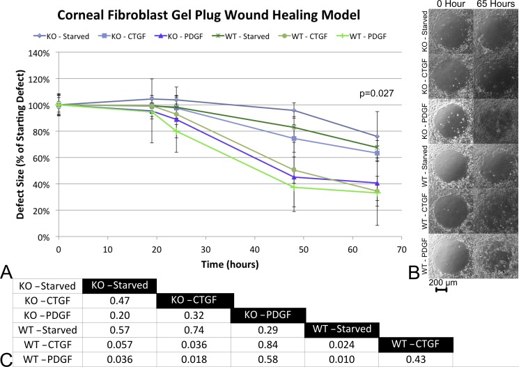
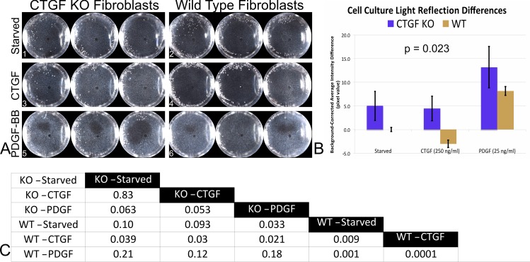
We sought to determine if connective tissue growth factor (CTGF) is necessary for the formation of corneal haze after corneal injury. Mice with post-natal, tamoxifen-induced, knockout of CTGF were subjected to excimer laser phototherapeutic keratectomy (PTK) and the corneas were allowed to heal. The extent of scaring was observed in non-induced mice, heterozygotes, and full homozygous knockout mice and quantified by macrophotography. The eyes from these mice were collected after euthanization for re-genotyping to control for possible Cre-mosaicism. Primary corneal fibroblasts from CTGF knockout corneas were established in a gel plug assay. The plug was removed, simulating an injury, and the rate of hole closure and the capacity for these cells to form light reflecting cells in response to CTGF and platelet-derived growth factor B (PDGF-B) were tested and compared to wild-type cells. We found that independent of genotype, each group of mice was still capable of forming light reflecting haze in the cornea after laser ablation (p = 0.40). Results from the gel plug closure rate in primary cell cultures of knockout cells were not statistically different from serum starved wild-type cells, independent of treatment. Compared to the serum starved wild-type cells, stimulation with PDGF-BB significantly increased the KO cell culture's light reflection (p = 0.03). Most interestingly, both reflective cultures were positive for α-SMA, but the cellular morphology and levels of α-SMA were distinct and not in proportion to the light reflection seen. This new work demonstrates that corneas without CTGF can still form sub-epithelial haze, and that the light reflecting phenotype can be reproduced in culture. These data support the possibilities of growth factor redundancy and that multiple pro-haze pathways exist.
Conflict of interest statement
Figures


Similar articles
-
A novel method for generating corneal haze in anterior stroma of the mouse eye with the excimer laser.Exp Eye Res. 2008 Feb;86(2):235-40. doi: 10.1016/j.exer.2007.10.014. Epub 2007 Nov 5. Exp Eye Res. 2008. PMID: 18068702 Free PMC article.
-
Growth factor expression in corneal wound healing after excimer laser keratectomy.Cornea. 1999 Sep;18(5):580-8. Cornea. 1999. PMID: 10487433
-
Conditional knockout of CTGF affects corneal wound healing.Invest Ophthalmol Vis Sci. 2014 Apr 1;55(4):2062-70. doi: 10.1167/iovs.13-12735. Invest Ophthalmol Vis Sci. 2014. PMID: 24627144 Free PMC article.
-
The corneal fibrosis response to epithelial-stromal injury.Exp Eye Res. 2016 Jan;142:110-8. doi: 10.1016/j.exer.2014.09.012. Exp Eye Res. 2016. PMID: 26675407 Free PMC article. Review.
-
Phototherapeutic keratectomy: 12 years of experience.Acta Ophthalmol Scand. 2003 Feb;81(1):19-32. doi: 10.1034/j.1600-0420.2003.00015.x. Acta Ophthalmol Scand. 2003. PMID: 12631015 Review.
Cited by
-
ECM stiffness modulates the proliferation but not the motility of primary corneal keratocytes in response to PDGF-BB.Exp Eye Res. 2022 Jul;220:109112. doi: 10.1016/j.exer.2022.109112. Epub 2022 May 18. Exp Eye Res. 2022. PMID: 35595094 Free PMC article.
-
Eyeing the Extracellular Matrix in Vascular Development and Microvascular Diseases and Bridging the Divide between Vascular Mechanics and Function.Int J Mol Sci. 2020 May 15;21(10):3487. doi: 10.3390/ijms21103487. Int J Mol Sci. 2020. PMID: 32429045 Free PMC article. Review.
References
-
- Blalock TD, Duncan MR, Varela JC, Goldstein MH, Tuli SS, Grotendorst GR, et al. Connective tissue growth factor expression and action in human corneal fibroblast cultures and rat corneas after photorefractive keratectomy. Invest Ophthalmol Vis Sci. 2003;44(5):1879–87. - PubMed
-
- Bradham DM, Igarashi A, Potter RL, Grotendorst GR. Connective tissue growth factor: a cysteine-rich mitogen secreted by human vascular endothelial cells is related to the SRC-induced immediate early gene product CEF-10. J Cell Biol. 1991;114(6):1285–94. PubMed Central PMCID: PMCPMC2289134. - PMC - PubMed
-
- Dammeier J, Brauchle M, Falk W, Grotendorst GR, Werner S. Connective tissue growth factor: a novel regulator of mucosal repair and fibrosis in inflammatory bowel disease? Int J Biochem Cell Biol. 1998;30(8):909–22. - PubMed
-
- Duncan MR, Frazier KS, Abramson S, Williams S, Klapper H, Huang X, et al. Connective tissue growth factor mediates transforming growth factor beta-induced collagen synthesis: down-regulation by cAMP. FASEB J. 1999;13(13):1774–86. - PubMed
MeSH terms
Substances
Grants and funding
LinkOut - more resources
Full Text Sources
Other Literature Sources
Medical
Molecular Biology Databases
Research Materials
Miscellaneous
