. 2017 Feb 16;12(2):e0172299.
doi: 10.1371/journal.pone.0172299. eCollection 2017.

Distinct susceptibility and applicability of MDCK derivatives for influenza virus research

Affiliations

Distinct susceptibility and applicability of MDCK derivatives for influenza virus research

Shih-Chao Lin et al. PLoS One. .

Abstract

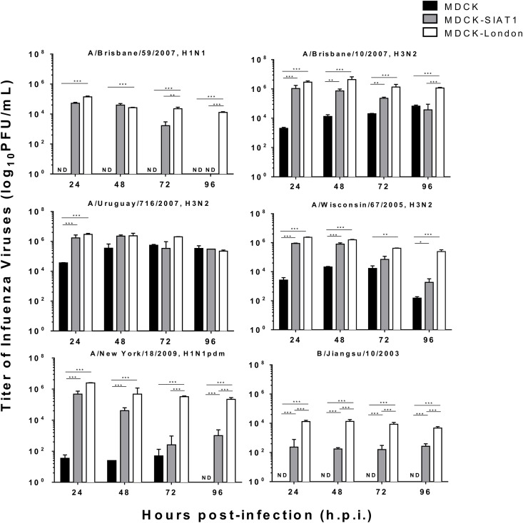
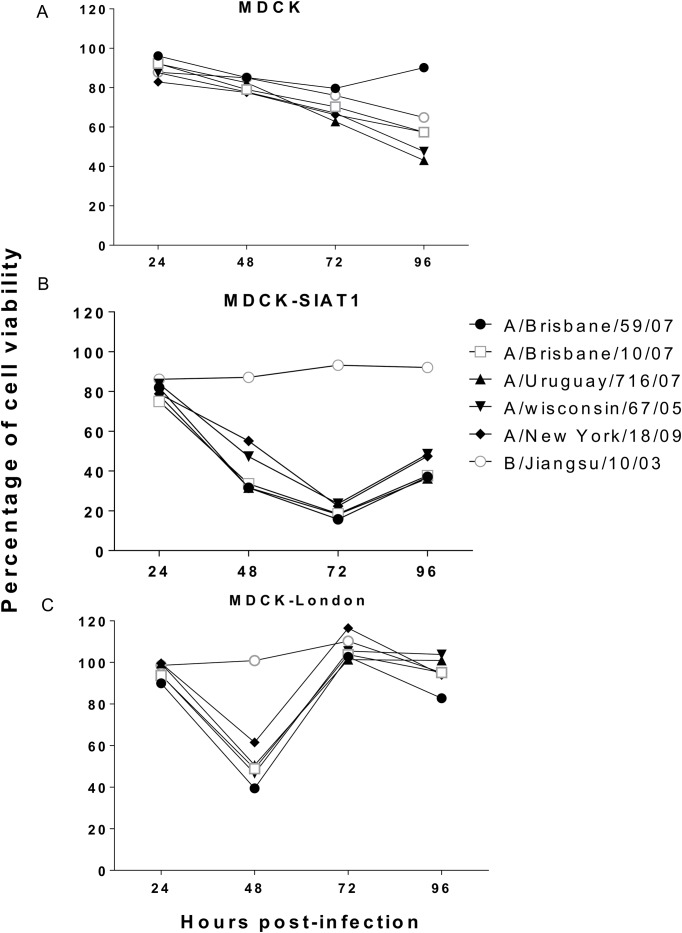
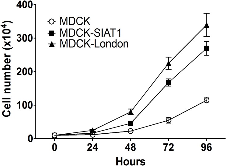
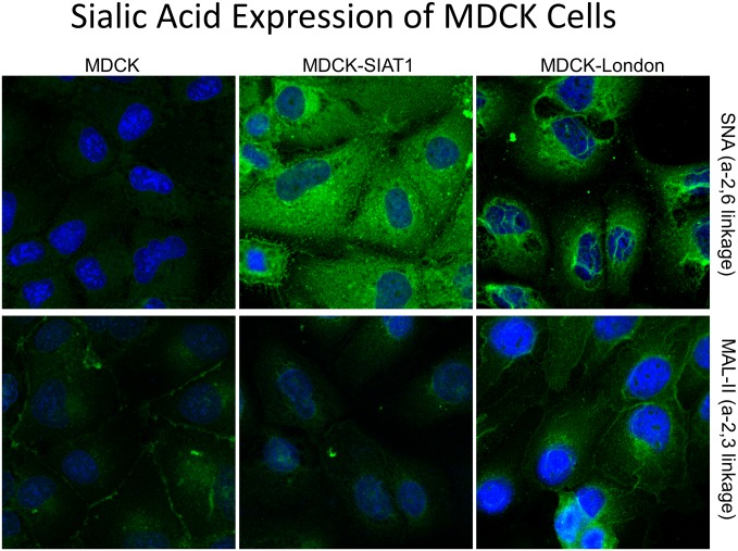
Madin-Darby Canine Kidney (MDCK) cells are widely utilized as a substrate for influenza virus isolation and propagation due to the high yields of virus. Here we compared the conventional MDCK cell line, MDCK-SIAT1 and MDCK-London for viral production, cell survival, and suitability in testing antivirals using six influenza strains including two H1N1 (pandemic and epidemic strains), three H3N2 and one influenza B strain. Overall our results suggest that MDCK-London cell line is superior for virus culturing and quantification, and hence an ideal platform to evaluate antiviral drug efficacy against multiple strains of influenza. Our data also suggests that while virus titers determined by the hemagglutination assay (HA) and neuraminidase activity (NA) are widely used to indicate viral load, there is a poor correlation between these measurements and the infectious titer obtained by plaque assay.


Conflict of interest statement

Competing Interests: The authors have declared that no competing interests exist.

Figures

Fig 1
Fig 1. Differential virus growth kinetics in three MDCK cell lines.
MDCK-lineage cell lines were infected βy epidemic H1N1 (A), pandemic H1N1 (B), H3N2 (C-E), and influenza B strain (F) with a M.O.I. of 0.01. Supernatant were collected at 24, 48, 72, and 96 hours post-infection. Viral titers were determined by Immunostaining Plaque Assay with MDCK-London cells. N.D. indicates Not Detectable. Data expressed as means ± SEM of three experiments in each group. *p <0.05, **P<0.01, ***p<0.001.
Fig 2
Fig 2. Cell viabilities of virus infected MDCK cells.
Cellular viability was determined as the concentration of formazan product level. MTS tetrazolium compound was added into influenza-infected MDCK (A), MDCK-SIAT1 (B), and MDCK-London (C) at 24, 48, 72, and 96 hours post-infection followed by incubating at 37°C for 1 hour. Concentration of formazan product was quantified with ELISA reader at the wavelength 490 nm. The baseline of viability from mock-infected cells (media only, no virus) was set to 100%. Data expressed as means ± SEM of three experiments in each group.
Fig 3
Fig 3. Cellular growth rates of MDCK-lineage cells.
Cells were seeded in plates with 10% FBS complete DMDM for 24 hours before counting numbers to allow adherence on the plates. The numbers of each three MDCK-lineage cells were counted by hemacytometer for at least 3 times in each independent experiment.
Fig 4
Fig 4. The expression and distribution of sialic acids in three MDCK-lineage cell lines.
Cells were seeded on chamber slides and stained with Sambucus nigra agglutinin (SNA, A-C) and Maackia amurensis agglutinin (MAL-II, D-F) lectins conjugated fluorescein after BSA blocking. Images were taken by confocal microscopy in a 680×magnification. Green fluorescence indicates the location of sialic acids and blue fluorescence stained by DAPI showed the nucleus of cells. Corrected total cell fluorescence (CTCF) was determined with software Image J (version 1.50i, Bethesda, MD USA). The highest density among three cell lines was defined as “high” whereas weakest was defined as “low”; density between high and low was considered “moderate”.

Similar articles

Cited by

References

    1. Hussain AI, Cordeiro M, Sevilla E, Liu J. Comparison of egg and high yielding MDCK cell-derived live attenuated influenza virus for commercial production of trivalent influenza vaccine: in vitro cell susceptibility and influenza virus replication kinetics in permissive and semi-permissive cells. Vaccine. 2010;28(22):3848–55. 10.1016/j.vaccine.2010.03.005 - DOI - PMC - PubMed
    1. Li IW, Chan KH, To KW, Wong SS, Ho PL, Lau SK, et al. Differential susceptibility of different cell lines to swine-origin influenza A H1N1, seasonal human influenza A H1N1, and avian influenza A H5N1 viruses. J Clin Virol. 2009;46(4):325–30. 10.1016/j.jcv.2009.09.013 - DOI - PubMed
    1. Zhai W, Zhang DN, Mai C, Choy J, Jian G, Sra K, et al. Comparison of different cell substrates on the measurement of human influenza virus neutralizing antibodies. PLoS One. 2012;7(12):e52327 10.1371/journal.pone.0052327 - DOI - PMC - PubMed
    1. Schepetiuk SK, Kok T. The use of MDCK, MEK and LLC-MK2 cell lines with enzyme immunoassay for the isolation of influenza and parainfluenza viruses from clinical specimens. J Virol Methods. 1993;42(2–3):241–50. - PubMed
    1. Govorkova EA, Murti G, Meignier B, de Taisne C, Webster RG. African green monkey kidney (Vero) cells provide an alternative host cell system for influenza A and B viruses. J Virol. 1996;70(8):5519–24. - PMC - PubMed

MeSH terms