. 2017 Mar 14;17(6):1116-1127.
doi: 10.1039/c6lc01165a.

Pneumatic stimulation of C. elegans mechanoreceptor neurons in a microfluidic trap

Affiliations

Pneumatic stimulation of C. elegans mechanoreceptor neurons in a microfluidic trap

Adam L Nekimken et al. Lab Chip. .

Abstract

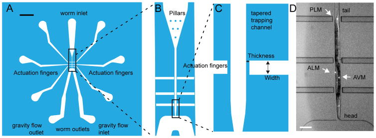
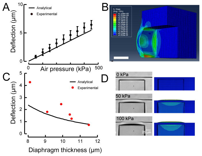
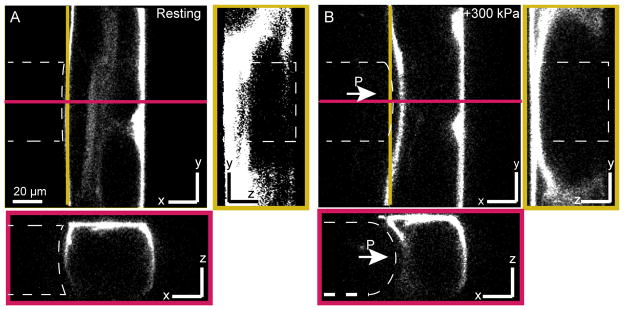
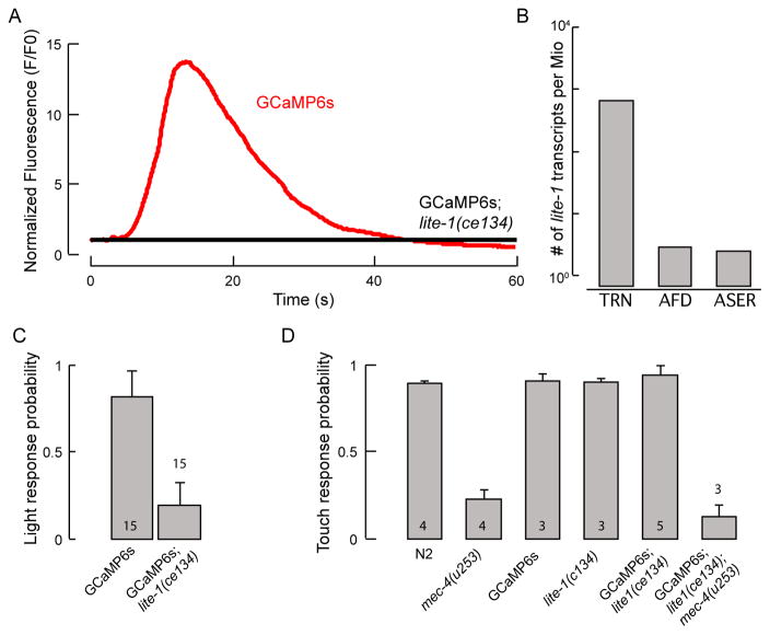
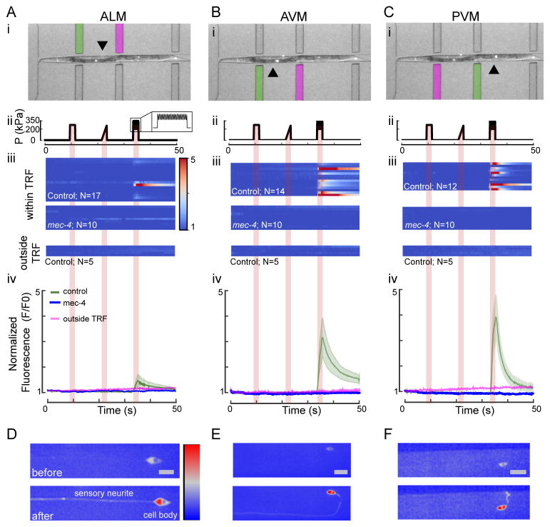
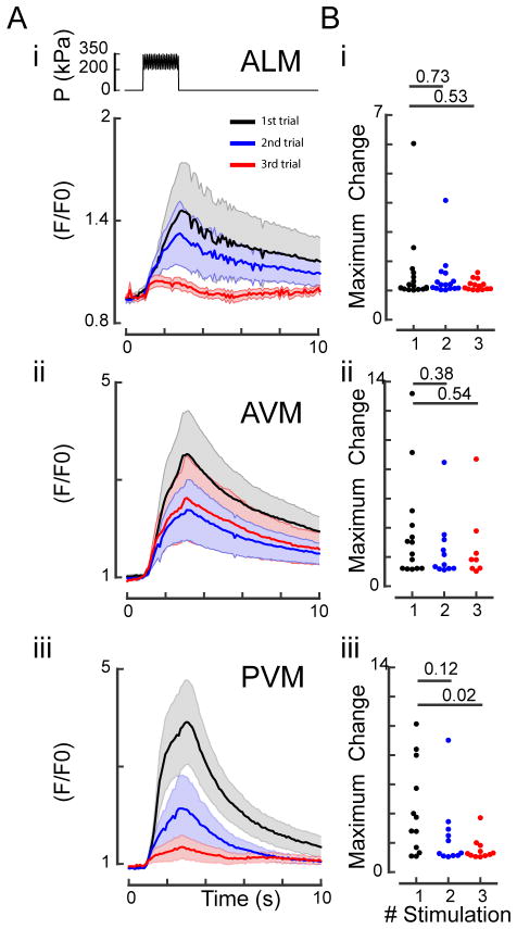
New tools for applying force to animals, tissues, and cells are critically needed in order to advance the field of mechanobiology, as few existing tools enable simultaneous imaging of tissue and cell deformation as well as cellular activity in live animals. Here, we introduce a novel microfluidic device that enables high-resolution optical imaging of cellular deformations and activity while applying precise mechanical stimuli to the surface of the worm's cuticle with a pneumatic pressure reservoir. To evaluate device performance, we compared analytical and numerical simulations conducted during the design process to empirical measurements made with fabricated devices. Leveraging the well-characterized touch receptor neurons (TRNs) with an optogenetic calcium indicator as a model mechanoreceptor neuron, we established that individual neurons can be stimulated and that the device can effectively deliver steps as well as more complex stimulus patterns. This microfluidic device is therefore a valuable platform for investigating the mechanobiology of living animals and their mechanosensitive neurons.


Figures

Fig. 1
Fig. 1. Channel design overview
A: Overall device design with inlet, outlet, and air reservoirs. Scale bar = 1 mm. B: Enlarged view of the pillars for worm orientation, the tapered trap, and the air channels. C: Enlarged view of two of the six independent pneumatic actuators with their diaphragms and the tip of the tapered trap, which is 24 μm in width at its smallest point. D: Representative micrograph of a trapped worm; Arrowheads point to the location of the TRN cell bodies. The mechanosensitive neurites extend anteriorly from the cell bodies toward the animal’s head. Scale bar = 100 μm.
Fig. 2
Fig. 2. Comparison of experimental measurements of diaphragm deflection to analytical and finite-element predictions
A: In the pressure range of interest, diaphragm deflection increases nearly linearly with applied pressure, in close agreement with the analytical simulations. Data are derived from measurements with a 10-μm-thick diaphragm and a 50-μm-wide actuator (black line). The variation in deflection could be attributed to variation in diaphragm thickness or material properties in the manufacturing process. B: Stress contour showing stress alleviation and stress concentration at PDMS-PDMS and PDMS-glass junctions respectively. Scale bar 20 μm. C: Experimental measurements of diaphragm deflection versus thickness for 100 kPa of applied pressure. Black solid line shows the analytical prediction. D: The shape of the deflected actuator in an experiment (left) matches the shape predicted by the finite element simulation (right).
Fig. 3
Fig. 3. Mechanical deformation of a live C. elegans in the microfluidic chip
Confocal slices through trapped animal stained with Dil in a 50×50-μm channel diaphragm before (A) and during (B) application of p=300 kPa to the pressure channel (outlined with a white dashed line). Left panels depict the yz-plane along the yellow line; the bottom panels show the xz-plane along the red line.
Fig. 4
Fig. 4. Light- and force-sensitive behaviors of C. elegans strains
A: A representative trace of TRN::GCaMP6s fluorescence intensity in the ALM TRN after illuminating a single worm with blue light in TRN::GCaMP6s control (red) and TRN::GCaMP6s;lite-1(ce314) mutants (black). N=10 worms have been imaged. Signals were normalized to the first 10 frames in after illumination. Note, difference in baseline might be due to calcium influx directly upon illumination with blue light. B: Quantification of lite-1(ce314) expression in PLM TRN by RNA-seq, in number of lite-1 mRNA reads found per million reads. C: Wild-type worms are more likely to show an avoidance response to blue light than lite-1(ce314) mutants when illuminated with 7 mW/mm2. Means and standard deviations are shown (N=15). D: Touch response of lite-1 and TRN::GCaMP6s transgenic animals. Mutants (TRN::GCaMP6s;lite-1(ce314)) and mec-4(u253), double mutants (TRN::GCaMP6s;lite-1(ce314)mec-4(u253)), and wild-type (N2) worms were assayed. Numbers in the bars indicate the number of independent experiments. In each experiment, 25 animals were touched 5 times with an eyebrow hair on the head and the tail in an alternating manner (10 touches in total per animal). Mean ± standard deviation.
Fig. 5
Fig. 5. Calcium dynamics of TRNs as a response to a step, ramp and buzz stimulus within and outside their presumptive tactile receptive fields (TRF)
A–C: Response dynamics of ALM (A), AVM (B) and PVM (C) touch receptor neurons stimulated within and outside their TRF. (i) Overlay of a brightfield and a fluorescence image showing a worm in the microfluidic chip. The actuator used for stimulation of the respective neuron within its receptive field is marked in green, and the actuator used to stimulate each neuron outside its TRF is marked in magenta. The arrows point toward the cell body of the neuron of interest. (ii) Stimulus protocol including 2 second diaphragm excitation representing a 275-kPa step, a 275-kPa ramp and a sine (75 kPa; 10 Hz) superimposed with a 275-kPa step (buzz). (iii) Multiple false color-coded normalized fluorescence intensity traces (F/F0) during the mechanical stimulation (shown in (ii)) of lite-1(ce314);TRN::GCaMP6s (control) and lite-1(ce314)mec-4(u253);TRN::GCaMP6s (mec-4) mutant animals and of control animals stimulated outside their TRF. (iv) Average fluorescence intensity (F/F0) of the traces shown in (ii) for control (mean ± SEM as green shaded area, N=17 for ALM, N=14 for AVM, N=12 for PVM) and mec-4 mutant animals (mean ± SEM in blue with N=10 animals for ALM, AVM, and PVM) when stimulated within their TRF and the average fluorescence intensity of five traces of each TRN when stimulated outside their TRF (magenta). The positive slope could be attributed to differential bleaching in the red and green channels. D–F: Representative high-resolution images of ALM, AVM and PVM neuron before and after stimulation within their TRF. Color scale = 1500–3500 gray values and scale bar (=10 μm) for all images.
Fig. 6
Fig. 6. Repeated mechanical stimulation of TRNs cause habituation of the calcium response
A: Response dynamics of ALM (i), AVM (ii) and PVM (iii) to three sequential buzz stimuli with an inter-stimulus interval of 1 min. Top panel in (i) shows the schematic representation of the stimulation protocol. A 75-kPa oscillation was superimposed to a 275-kPa step for 2 s. Normalized TRN::GCaMP6s fluorescence for three sequential stimuli. Black traces, fluorescence during the first stimulus; blue traces, fluorescence during the second stimulus; red traces, fluorescence during the third stimulus. Means and standard error of the mean (N=10) are shown. B: Peak amplitude after sequential stimuli. Numbers above individual data points represent P-values according to Wilcoxon test for statistical significance.

Similar articles

Cited by

References

    1. Hughes S, McBain S, Dobson J, El Haj AJ. Journal of The Royal Society Interface. 2008;5:855–863. - PMC - PubMed
    1. Sachs F. Physiology. 2010;25:50–56. - PMC - PubMed
    1. Chen TW, Wardill TJ, Sun Y, Pulver SR, Renninger SL, Baohan A, Schreiter ER, Kerr RA, Orger MB, Jayaraman V, Looger LL, Svoboda K, Kim DS. Nature. 2013;499:295–300. - PMC - PubMed
    1. Reits EA, Neefjes JJ. Nature Cell Biology. 2001;3:E145–E147. - PubMed
    1. Krieg M, Dunn AR, Goodman MB. Nature Cell Biology. 2014;16:224–233. - PMC - PubMed

Publication types

MeSH terms