

Involuntary eye motion correction in retinal optical coherence tomography: Hardware or software solution?
- PMID: 28208100
- PMCID: PMC5350634
- DOI: 10.1016/j.media.2017.02.002
Involuntary eye motion correction in retinal optical coherence tomography: Hardware or software solution?
Abstract
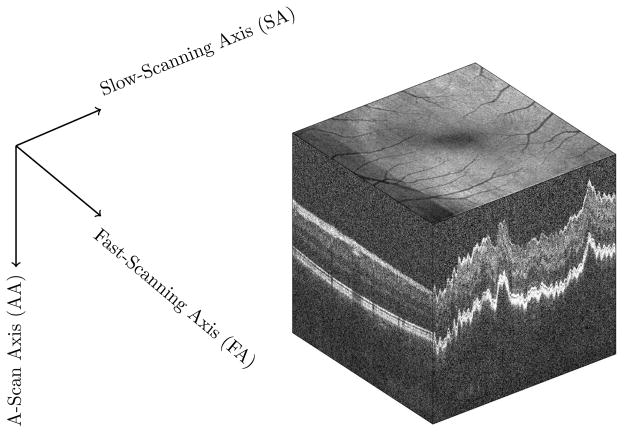
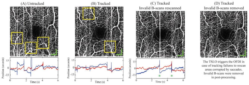
In this paper, we review state-of-the-art techniques to correct eye motion artifacts in Optical Coherence Tomography (OCT) imaging. The methods for eye motion artifact reduction can be categorized into two major classes: (1) hardware-based techniques and (2) software-based techniques. In the first class, additional hardware is mounted onto the OCT scanner to gather information about the eye motion patterns during OCT data acquisition. This information is later processed and applied to the OCT data for creating an anatomically correct representation of the retina, either in an offline or online manner. In software based techniques, the motion patterns are approximated either by comparing the acquired data to a reference image, or by considering some prior assumptions about the nature of the eye motion. Careful investigations done on the most common methods in the field provides invaluable insight regarding future directions of the research in this area. The challenge in hardware-based techniques lies in the implementation aspects of particular devices. However, the results of these techniques are superior to those obtained from software-based techniques because they are capable of capturing secondary data related to eye motion during OCT acquisition. Software-based techniques on the other hand, achieve moderate success and their performance is highly dependent on the quality of the OCT data in terms of the amount of motion artifacts contained in them. However, they are still relevant to the field since they are the sole class of techniques with the ability to be applied to legacy data acquired using systems that do not have extra hardware to track eye motion.
Keywords: Motion artifact correction; Optical Coherence Tomography (OCT); Retina.
Copyright © 2017 Elsevier B.V. All rights reserved.
Figures


Similar articles
-
Motion artefact correction in retinal optical coherence tomography using local symmetry.Med Image Comput Comput Assist Interv. 2014;17(Pt 2):130-7. doi: 10.1007/978-3-319-10470-6_17. Med Image Comput Comput Assist Interv. 2014. PMID: 25485371
-
Bright-Field Imaging and Optical Coherence Tomography of the Mouse Posterior Eye.Methods Mol Biol. 2016;1438:395-415. doi: 10.1007/978-1-4939-3661-8_20. Methods Mol Biol. 2016. PMID: 27150100
-
Impact of eye-tracking technology on OCT-angiography imaging quality in age-related macular degeneration.Graefes Arch Clin Exp Ophthalmol. 2017 Aug;255(8):1535-1542. doi: 10.1007/s00417-017-3684-z. Epub 2017 May 4. Graefes Arch Clin Exp Ophthalmol. 2017. PMID: 28474129 Clinical Trial.
-
Recent advances in wide field and ultrawide field optical coherence tomography angiography in retinochoroidal pathologies.Expert Rev Med Devices. 2021 Apr;18(4):375-386. doi: 10.1080/17434440.2021.1902301. Epub 2021 Mar 23. Expert Rev Med Devices. 2021. PMID: 33724126 Review.
-
Heidelberg Spectralis Optical Coherence Tomography Angiography: Technical Aspects.Dev Ophthalmol. 2016;56:1-5. doi: 10.1159/000442768. Epub 2016 Mar 15. Dev Ophthalmol. 2016. PMID: 27022921 Review.
Cited by
-
Numerical method for axial motion artifact correction in retinal spectral-domain optical coherence tomography.Front Optoelectron. 2020 Dec;13(4):393-401. doi: 10.1007/s12200-019-0951-0. Epub 2019 Dec 15. Front Optoelectron. 2020. PMID: 36641561 Free PMC article.
-
In-vivo 3D corneal elasticity using air-coupled ultrasound optical coherence elastography.Biomed Opt Express. 2019 Nov 14;10(12):6272-6285. doi: 10.1364/BOE.10.006272. eCollection 2019 Dec 1. Biomed Opt Express. 2019. PMID: 31853399 Free PMC article.
-
Enhancing OCT Reliability: The Role of Eye-Tracking in Achieving Consistent Retinal Measurements [Response to Letter].Clin Ophthalmol. 2024 Dec 3;18:3579-3580. doi: 10.2147/OPTH.S505608. eCollection 2024. Clin Ophthalmol. 2024. PMID: 39649983 Free PMC article. No abstract available.
-
LEARNING TO CORRECT AXIAL MOTION IN OCT FOR 3D RETINAL IMAGING.Proc Int Conf Image Proc. 2021 Sep;2021:126-130. doi: 10.1109/icip42928.2021.9506620. Epub 2021 Aug 23. Proc Int Conf Image Proc. 2021. PMID: 35950046 Free PMC article.
-
Spiral scanning improves subject fixation in widefield retinal imaging.Opt Lett. 2024 May 1;49(9):2489-2492. doi: 10.1364/OL.517088. Opt Lett. 2024. PMID: 38691751 Free PMC article.
References
-
- Adler DC, Ko TH, Fujimoto JG. Speckle reduction in optical coherence tomography images by use of a spatially adaptive wavelet filter. Optics letters. 2004;29(24):2878–2880. - PubMed
-
- Alonso-Caneiro D, Read SA, Collins MJ. Speckle reduction in optical coherence tomography imaging by affine-motion image registration. Journal of biomedical optics. 2011;16(11):116027–1160275. - PubMed
-
- Avanaki MR, Cernat R, Tadrous PJ, Tatla T, Podoleanu AG, Hojjatoleslami SA. Spatial compounding algorithm for speckle reduction of dynamic focus oct images. Photonics Technology Letters, IEEE. 2013;25(15):1439–1442.
-
- Baghaie A, D’souza RM, Yu Z. Sparse and low rank decomposition based batch image alignment for speckle reduction of retinal oct images. Biomedical Imaging (ISBI), 2015 IEEE 12th International Symposium on; IEEE; 2015a. pp. 226–230.
Publication types
MeSH terms
Grants and funding
LinkOut - more resources
Full Text Sources
Other Literature Sources
