Review
. 2017 Feb 11;18(2):382.
doi: 10.3390/ijms18020382.

Endoplasmic Reticulum (ER) Stress and Endocrine Disorders

Affiliations
Review

Endoplasmic Reticulum (ER) Stress and Endocrine Disorders

Daisuke Ariyasu et al. Int J Mol Sci. .

Abstract

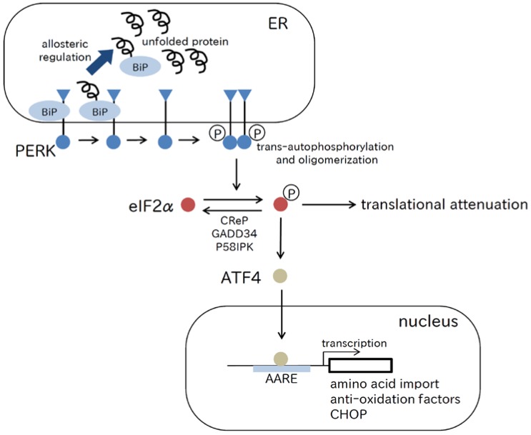
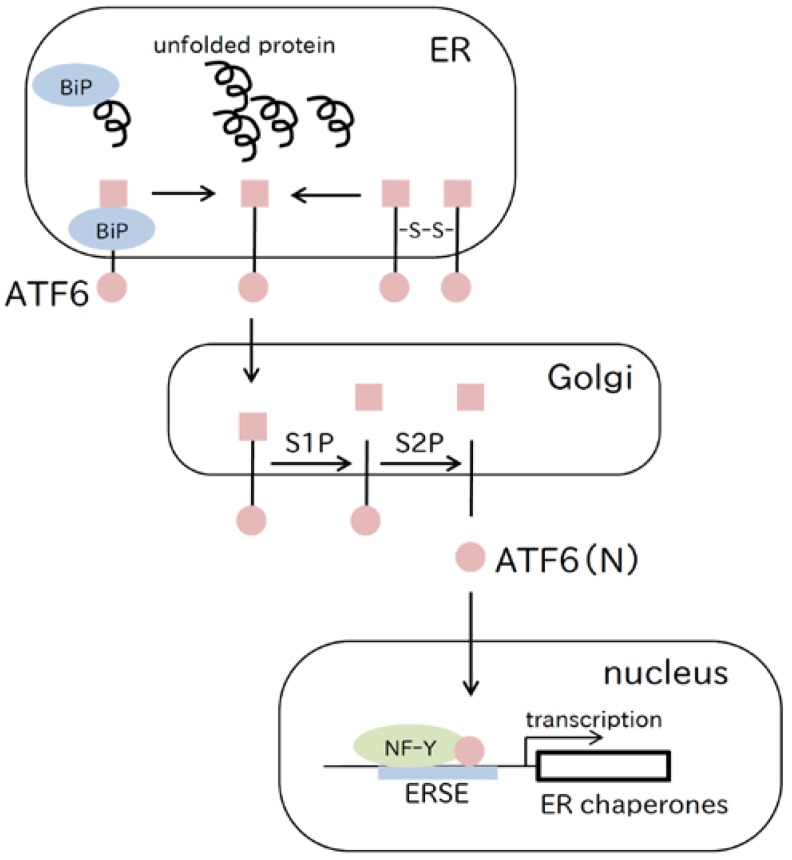
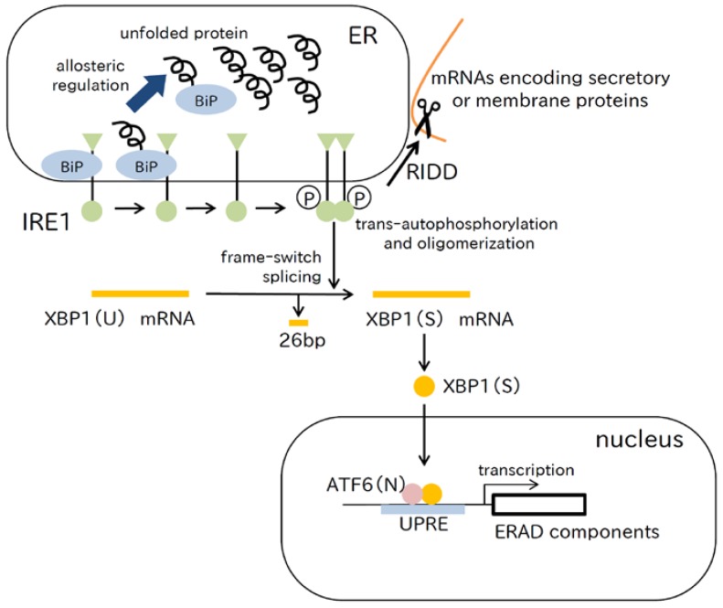
The endoplasmic reticulum (ER) is the organelle where secretory and membrane proteins are synthesized and folded. Unfolded proteins that are retained within the ER can cause ER stress. Eukaryotic cells have a defense system called the "unfolded protein response" (UPR), which protects cells from ER stress. Cells undergo apoptosis when ER stress exceeds the capacity of the UPR, which has been revealed to cause human diseases. Although neurodegenerative diseases are well-known ER stress-related diseases, it has been discovered that endocrine diseases are also related to ER stress. In this review, we focus on ER stress-related human endocrine disorders. In addition to diabetes mellitus, which is well characterized, several relatively rare genetic disorders such as familial neurohypophyseal diabetes insipidus (FNDI), Wolfram syndrome, and isolated growth hormone deficiency type II (IGHD2) are discussed in this article.

Keywords: PKR-like endoplasmic reticulum kinase (PERK); activating transcription factor 6 (ATF6); chemical chaperone; endocrine disorder; endoplasmic reticulum stress; inositol requirement 1 (IRE1); old astrocyte specifically induced substance (OASIS) family.


Conflict of interest statement

The authors declare no conflict of interest.

Figures

Figure 1
Figure 1
The PERK pathway. PERK oligomerizes and trans-autophosphorylates by detecting unfolded proteins in the ER, inactivates eIF2α by phosphorylation, and induces translation of ATF4. According to the current allosteric model of UPR induction, the ATPase domain of BiP interacts with the luminal domain of PERK that dissociates when an unfolded protein binds to the canonical substrate binding domain of BiP. ATF4 enhances transcription of genes involved in amino acid transport, oxidative stress, and apoptosis. Phosphorylated eIF2α is dephosphorylated by CReP, GADD34, and P58IPK. AARE, amino acid response element; ATF4, activating transcription factor 4; CHOP, C/EBP-homologous protein; CReP, constitutive repressor of eIF2α phosphorylation; eIF2α, eukaryotic translational initiation factor 2; GADD34, growth arrest and DNA damage 34; PERK, PKR-like endoplasmic reticulum kinase; P58IPK, 58 kDa-inhibitor of protein kinase.
Figure 2
Figure 2
The ATF6 pathway. ATF6 detects unfolded proteins in the ER, and translocates to the Golgi apparatus. In the Golgi, ATF6 is cleaved by site 1 protease (S1P) and site 2 protease (S2P), and the N-terminal portion, ATF6(N), is translocated to the nucleus. ATF6(N) binds to the ERSE forming a heterodimer with NF-Y and enhances transcription of ER chaperone genes.
Figure 3
Figure 3
The IRE1 pathway. Using the allosteric model of UPR induction, the mechanism of detecting unfolded proteins by the luminal domain of IRE1 is shown. The ATPase domain of BiP interacts with the luminal domain of IRE1, which dissociates when an unfolded protein binds to the canonical substrate binding domain of BiP. IRE1 is activated by oligomerization and trans-autophosphorylation. Next, IRE1 converts XBP1(U) mRNA into XBP1(S) mRNA by frame-switch splicing, which leads to XBP1(S) production. XBP1(S) translocates to the nucleus, and binds to the UPRE forming a heterodimer with ATF6(N), which results in enhancement of gene expression encoding ERAD components. IRE1 RNase activity degrades mRNAs associated with the ER membrane, thereby unburdening the protein load through regulated IRE1-dependent decay of mRNA (RIDD).

References

    1. Ahner A., Brodsky J.L. Checkpoints in ER-associated degradation: Excuse me, which way to the proteasome? Trends Cell Biol. 2004;14:474–478. doi: 10.1016/j.tcb.2004.07.013. - DOI - PubMed
    1. Yoshida H. ER stress and diseases. FEBS J. 2007;274:630–658. doi: 10.1111/j.1742-4658.2007.05639.x. - DOI - PubMed
    1. Katayama T., Imaizumi K., Sato N., Miyoshi K., Kudo T., Hitomi J., Morihara T., Yoneda T., Gomi F., Mori Y., et al. Presenilin-1 mutations downregulate the signalling pathway of the unfolded-protein response. Nat. Cell Biol. 1999;1:479–485. doi: 10.1038/70265. - DOI - PubMed
    1. Shimura H., Hattori N., Kubo S., Mizuno Y., Asakawa S., Minoshima S., Shimizu N., Iwai K., Chiba T., Tanaka K., et al. Familial Parkinson disease gene product, parkin, is a ubiquitin-protein ligase. Nat. Genet. 2000;25:302–305. - PubMed
    1. Schaffar G., Breuer P., Boteva R., Behrends C., Tzvetkov N., Strippel N., Sakahira H., Siegers K., Hayer-Hartl M., Hartl F.U. Cellular toxicity of polyglutamine expansion proteins: Mechanism of transcription factor deactivation. Mol. Cell. 2004;15:95–105. doi: 10.1016/j.molcel.2004.06.029. - DOI - PubMed

LinkOut - more resources