Review
. 2017 Feb 8;6(1):11.
doi: 10.3390/biology6010011.

The Glutamate Dehydrogenase Pathway and Its Roles in Cell and Tissue Biology in Health and Disease

Affiliations
Review

The Glutamate Dehydrogenase Pathway and Its Roles in Cell and Tissue Biology in Health and Disease

Andreas Plaitakis et al. Biology (Basel). .

Abstract

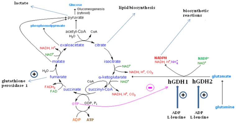
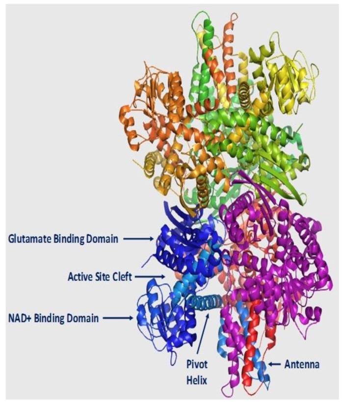
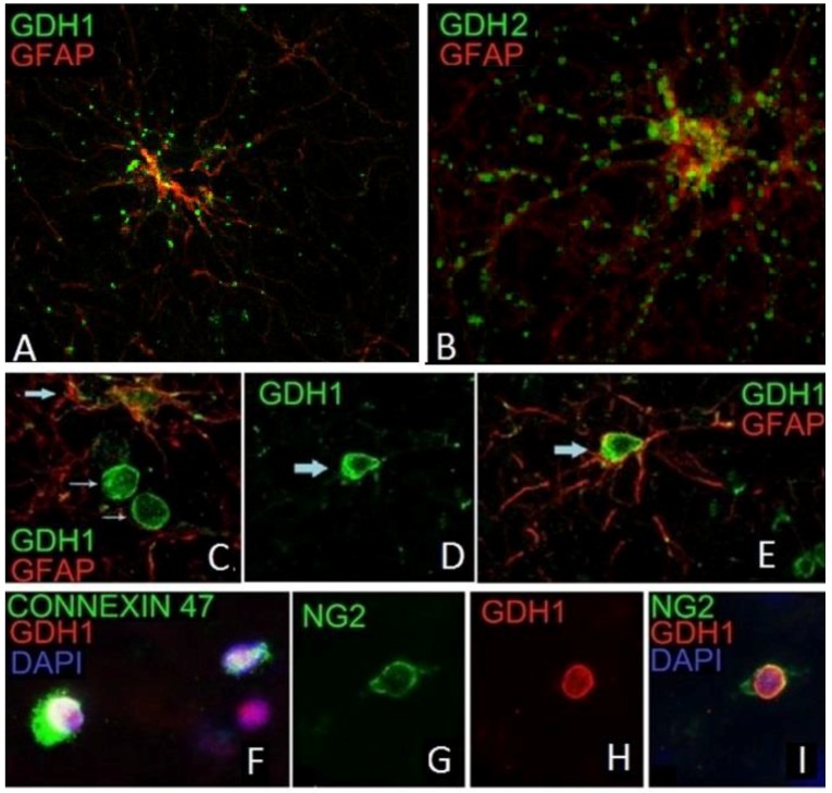
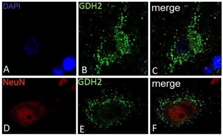
Glutamate dehydrogenase (GDH) is a hexameric enzyme that catalyzes the reversible conversion of glutamate to α-ketoglutarate and ammonia while reducing NAD(P)⁺ to NAD(P)H. It is found in all living organisms serving both catabolic and anabolic reactions. In mammalian tissues, oxidative deamination of glutamate via GDH generates α-ketoglutarate, which is metabolized by the Krebs cycle, leading to the synthesis of ATP. In addition, the GDH pathway is linked to diverse cellular processes, including ammonia metabolism, acid-base equilibrium, redox homeostasis (via formation of fumarate), lipid biosynthesis (via oxidative generation of citrate), and lactate production. While most mammals possess a single GDH1 protein (hGDH1 in the human) that is highly expressed in the liver, humans and other primates have acquired, via duplication, an hGDH2 isoenzyme with distinct functional properties and tissue expression profile. The novel hGDH2 underwent rapid evolutionary adaptation, acquiring unique properties that enable enhanced enzyme function under conditions inhibitory to its ancestor hGDH1. These are thought to provide a biological advantage to humans with hGDH2 evolution occurring concomitantly with human brain development. hGDH2 is co-expressed with hGDH1 in human brain, kidney, testis and steroidogenic organs, but not in the liver. In human cerebral cortex, hGDH1 and hGDH2 are expressed in astrocytes, the cells responsible for removing and metabolizing transmitter glutamate, and for supplying neurons with glutamine and lactate. In human testis, hGDH2 (but not hGDH1) is densely expressed in the Sertoli cells, known to provide the spermatids with lactate and other nutrients. In steroid producing cells, hGDH1/2 is thought to generate reducing equivalents (NADPH) in the mitochondria for the biosynthesis of steroidal hormones. Lastly, up-regulation of hGDH1/2 expression occurs in cancer, permitting neoplastic cells to utilize glutamine/glutamate for their growth. In addition, deregulation of hGDH1/2 is implicated in the pathogenesis of several human disorders.

Keywords: GDH; GDH deregulation and diseases; expression; glioma; hGDH1; hGDH2; human tissues; regulation; structure.


Conflict of interest statement

The authors declare no conflict of interest.

Figures

Figure 1
Figure 1
The glutamate dehydrogenase (GDH) pathway and the Krebs cycle function. As shown here, oxidative deamination of glutamate by hGDH1 and hGDH2 generates α-ketoglutarate, ammonia and NADH or NADPH. While α-ketoglutarate is metabolized by the Krebs cycle, NADPH can be used for biosynthetic reactions. GTP generated at the step of succinyl-CoA to succinate potently inhibits hGDH1 thus preventing glutamate from fueling this cycle under an adequate energy charge. On the other hand, hGDH2 has dissociated its function from GTP control and as such it permits glutamate flux under conditions inhibitory to hGDH1. Both enzymes are activated by ADP and L-leucine, which can act synergistically. Oxidative metabolism of α-ketoglutarate via the Krebs cycle generates citrate that can support the biosynthesis of lipids required for nerve tissue development and/or glioma cell growth. Also, it can generate lactate to be provided by astrocytes to neurons or by Sertoli cells to spermatids. Fumarate, generated by this pathway, is shown to stimulate glutathione peroxidase 1 activity, thus contributing to homeostasis against oxidative stress This Figure is modified from [7].
Figure 2
Figure 2
Structural model of hGDH1. Shown is a cartoon diagram of the apo form of the hGDH1 hexamer (PDB code: 1L1F), where each of the six subunits is colored differently. The main parts of the GDH subunit (active site, NAD+ binding domain, glutamate binding domain, antenna and pivot helix) are indicated only for the blue subunit. The figure was constructed using the PyMOL Molecular Graphics System, Version 1.8 (Schrodinger LLC., Cambridge, MA, USA).
Figure 3
Figure 3
hGDH1 and hGDH2 expression in glial cells. Punctate immunoreactivity for hGDH1 (A) and hGDH2 (B) is present in the cytoplasm and along the proximal and distal processes of GFAP positive astrocytes (IF images of high magnification). Also, hGDH1-specific staining is detected in the nucleus of a GFAP positive astrocyte (C,D,E), of a connexin 47-positive oligodendrocyte (F) and of an NG2-positive oligodendrocyte-precursor (G,H,I) (IF images of unfixed human frontal lobe cortex. Blue staining represents DAPI labeled nuclei).
Figure 4
Figure 4
Localization of hGDH2-positive “puncta” in the periphery and in the perinuclear cytoplasmic area of large human cortical neurons. Specific hGDH2 punctate staining is observed in the cytoplasm of a large neuron from the occipital lobe within coarse structures resembling mitochondria (AC). A constellation of intense hGDH2-specific “puncta” is also detected in the periphery of a sizable frontal lobe neuron with a large nucleus and prominent nucleolus (DF). (IF images of unfixed human frontal and occipital lobe cortex. Blue staining represents DAPI labeled cell nuclei).
Figure 5
Figure 5
Association of hGDH2 with the nuclear membrane in small cortical neurons. A delicate circular GDH2 specific-staining is detected around the NeuN-positive area (AC). Double IF using anti-hGDH2 and anti lamin A/C specific antiserums revealed that hGDH2 co-localizes with Lamin A/C on the nuclear membrane of this cell (DF). (IF images of unfixed human frontal lobe cortex).

References

    1. Smith E.L., Austin B.M., Blumenthal K.M., Nyc J.F. In: The Enzymes. Boyer P.D., editor. Academic Press; New York, NY, USA: 1975. pp. 293–367.
    1. Duran R.V., Oppliger W., Robitaille A.M., Heiserich L., Skendaj R., Gottlieb E., Hall M.N. Glutaminolysis activates Rag-mTORC1 signaling. Mol. Cell. 2012;47:349–358. doi: 10.1016/j.molcel.2012.05.043. - DOI - PubMed
    1. Jin L., Li D., Alesi G.N., Fan J., Kang H.B., Lu Z., Boggon T.J., Jin P., Yi H., Wright E.R., et al. Glutamate dehydrogenase 1 signals through antioxidant glutathione peroxidase 1 to regulate redox homeostasis and tumor growth. Cancer Cell. 2015;27:257–270. doi: 10.1016/j.ccell.2014.12.006. - DOI - PMC - PubMed
    1. Engel P.C. Glutamate Dehydrogenases: The Why and How of Coenzyme Specificity. Neurochem. Res. 2014;39:426–432. doi: 10.1007/s11064-013-1089-x. - DOI - PubMed
    1. Miller S.M., Magasanik B. Role of NAD-linked glutamate dehydrogenase in nitrogen metabolism in Saccharomyces cerevisiae. J. Bacteriol. 1990;172:4927–4935. doi: 10.1128/jb.172.9.4927-4935.1990. - DOI - PMC - PubMed

LinkOut - more resources