Review
. 2017 Feb 8;8(2):62.
doi: 10.3390/genes8020062.

Elaborated Action of the Human Primosome

Affiliations
Review

Elaborated Action of the Human Primosome

Andrey G Baranovskiy et al. Genes (Basel). .

Abstract

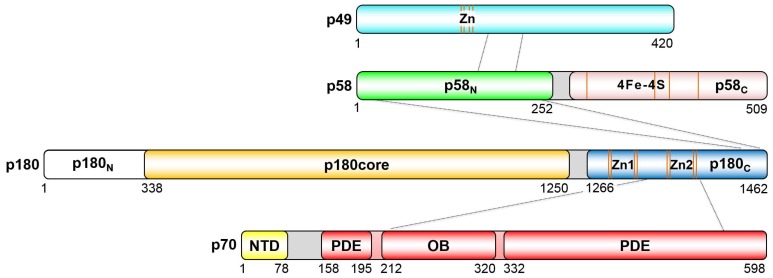
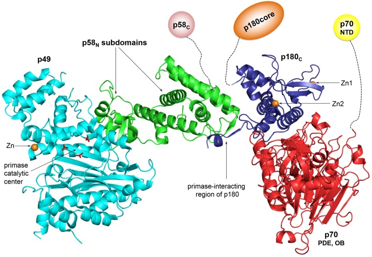
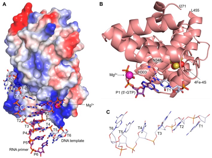
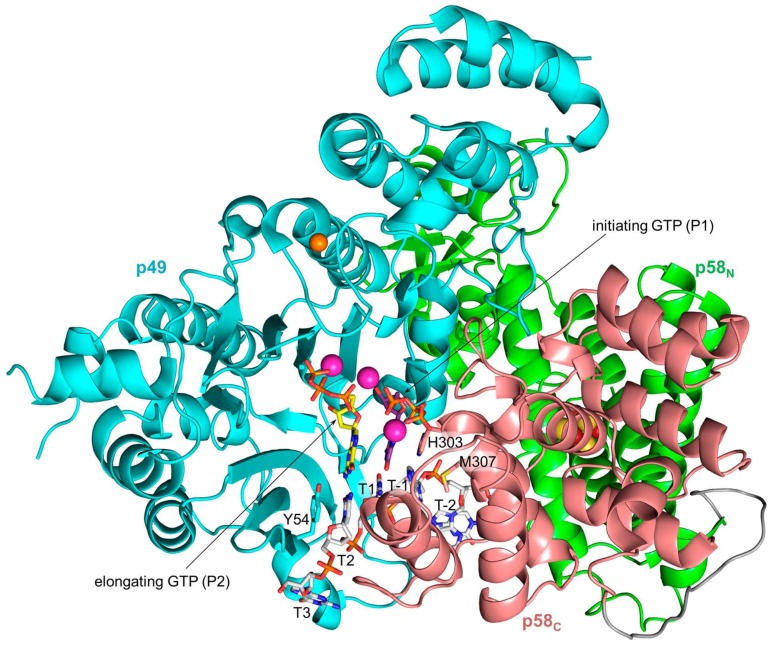
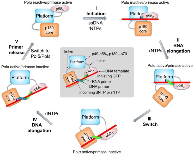
The human primosome is a 340-kilodalton complex of primase (DNA-dependent RNA polymerase) and DNA polymerase α, which initiates genome replication by synthesizing chimeric RNA-DNA primers for DNA polymerases δ and ϵ. Accumulated biochemical and structural data reveal the complex mechanism of concerted primer synthesis by two catalytic centers. First, primase generates an RNA primer through three steps: initiation, consisting of dinucleotide synthesis from two nucleotide triphosphates; elongation, resulting in dinucleotide extension; and termination, owing to primase inhibition by a mature 9-mer primer. Then Polα, which works equally well on DNA:RNA and DNA:DNA double helices, intramolecularly catches the template primed by a 9mer RNA and extends the primer with dNTPs. All primosome transactions are highly coordinated by autoregulation through the alternating activation/inhibition of the catalytic centers. This coordination is mediated by the small C-terminal domain of the primase accessory subunit, which forms a tight complex with the template:primer, shuttles between the primase and DNA polymerase active sites, and determines their access to the substrate.

Keywords: DNA polymerase α; DNA replication; RNA synthesis; human; initiation; primase; primosome; protein-DNA interaction; steric hindrance; termination.


Conflict of interest statement

The authors declare no conflicts of interest.

Figures

Figure 1
Figure 1
Schematic representation of the domain organization in the human primosome. The borders of the regions participating in intersubunit interactions are designated by dotted lines. Positions of the conserved cysteines coordinating zinc or [4Fe-4S] cluster are indicated by orange lines. The linkers responsible for flexible connections between domains are colored gray.
Figure 2
Figure 2
The platform of the human primosome. Coordinates of the human primosome (PDB ID 5EXR) were used to represent the platform structure. The color scheme for domains is the same as in Figure 1. The positions of p58C and p180core, as well as the linkers connecting them to the platform, vary depending on the primer synthesis step. For space-saving purposes, p58C, p180core, and p70-NTD are shown at reduced scale relative to the platform. All figures were prepared using the PyMOL Molecular Graphics System (version 1.8, Schrödinger, LLC).
Figure 3
Figure 3
Interaction of p58C with a DNA template primed by RNA. (A) p58C specifically recognizes the DNA:RNA junction at the primer 5′-end containing the triphosphate. The p58C surface is represented by the vacuum electrostatic potential at 20% transparency; (B) mechanism of p58C specificity to a purine at the initiation site. The hydrogen bond is depicted by dashed blue line; (C) DNA template bends between T3 and T4. All parts of the figure were drawn using the coordinates of the p58C/DNA:RNA complex (PDB ID 5F0Q).
Figure 4
Figure 4
The model of human primase in the initiation complex with a DNA template and two GTP molecules. The linker between p58N and p58C colored gray is shown for reference purposes only. The carbons of the DNA template, initiating GTP, and elongating GTP are colored gray, purple, and yellow, respectively. The atoms of zinc, magnesium, iron, and sulfur are represented as spheres and colored orange, magenta, red, and yellow, respectively.
Figure 5
Figure 5
Interaction of human primase with a DNA template and rNTPs during RNA synthesis initiation. The color scheme is the same as in Figure 4. The residues of p49 interacting with the DNA template and the initiating GTP are identified from the model of the initiation complex. The asterisk indicates that a main-chain atom of the amino acid forms a hydrogen bond with a nucleotide. Amino acids participating in stacking interactions with nucleotides are shown in rectangular boxes. Interactions of aspartates 109 and 111 with both rNTPs are mediated by the Mg2+ ions.
Figure 6
Figure 6
The model of human primase in elongation complex with a DNA template, primed by 7-mer RNA, and an incoming GTP. The curved arrow shows the direction of p58C rotation relative to p49-p58N during primer extension. The atoms of zinc, magnesium, iron, and sulfur are represented as spheres and colored orange, magenta, red, and yellow, respectively.
Figure 7
Figure 7
The model of the switch complex containing p180core, p58C, a DNA template primed by a 9-mer RNA, and incoming dCTP. p180core subdomains are shown in different colors. The carbons of the DNA template, RNA primer, and incoming dCTP are colored gray, purple, and yellow, respectively. This model was made using the coordinates of the p180core/DNA:RNA/dCTP complex (PDB ID 4QCL) and p58C/DNA:RNA complex (PDB ID 5F0Q).
Figure 8
Figure 8
Close-up view of the Polα active site with bound aphidicolin and the DNA:RNA duplex. The color scheme for p180core subdomains is same as in Figure 7. The carbons of aphidicolin are colored wheat. Side chains of the key residues, participating in hydrophobic interactions with aphidicolin and in stabilization of the syn conformation of the templating guanine, are shown as sticks. RNA primer contains a dideoxy-cytidine at the 3′-end. This figure was drawn using the coordinates of the p180core/DNA:RNA/aphidicolin complex (PDB ID 4Q5V).
Figure 9
Figure 9
Schematic representation of conformational changes in the primosome during chimeric primer synthesis. At the first step (steps are labeled by roman numerals), p58C moves toward p49 to initiate RNA synthesis. During the second step, p58C moves toward p180core and pushes it to dissociate from the platform. Additionally, when RNA primer length is nine nucleotides, p58C makes a steric hindrance with the platform, which prevents primer extension by p49. At the third step, p58C rotates and loads the template:primer to the Polα active site. At the fourth step, Polα extends the RNA primer with dNTPs. At the fifth step, the primosome is replaced by Polε or Polδ.

References

    1. Muzi-Falconi M., Giannattasio M., Foiani M., Plevani P. The DNA polymerase α-primase complex: Multiple functions and interactions. Sci. World J. 2003;3:21–33. doi: 10.1100/tsw.2003.05. - DOI - PMC - PubMed
    1. Pavlov Y.I., Shcherbakova P.V. DNA polymerases at the eukaryotic fork-20 years later. Mutat. Res. 2010;685:45–53. doi: 10.1016/j.mrfmmm.2009.08.002. - DOI - PMC - PubMed
    1. Lujan S.A., Williams J.S., Kunkel T.A. DNA polymerases divide the labor of genome replication. Trends Cell Biol. 2016;26:640–654. doi: 10.1016/j.tcb.2016.04.012. - DOI - PMC - PubMed
    1. Smith D.J., Whitehouse I. Intrinsic coupling of lagging-strand synthesis to chromatin assembly. Nature. 2012;483:434–438. doi: 10.1038/nature10895. - DOI - PMC - PubMed
    1. Pellegrini L. The pol α-primase complex. Subcell. Biochem. 2012;62:157–169. - PubMed

LinkOut - more resources