. 2017 Feb 9;18(2):77.
doi: 10.3390/ijms18020077.

Gypenoside XVII Prevents Atherosclerosis by Attenuating Endothelial Apoptosis and Oxidative Stress: Insight into the ERα-Mediated PI3K/Akt Pathway

Affiliations

Gypenoside XVII Prevents Atherosclerosis by Attenuating Endothelial Apoptosis and Oxidative Stress: Insight into the ERα-Mediated PI3K/Akt Pathway

Ke Yang et al. Int J Mol Sci. .

Abstract

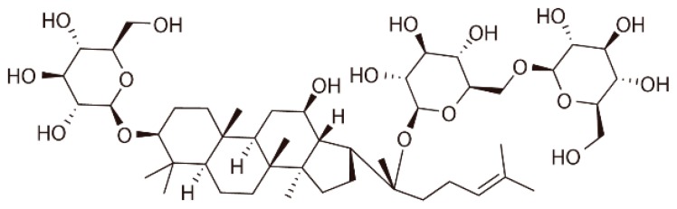
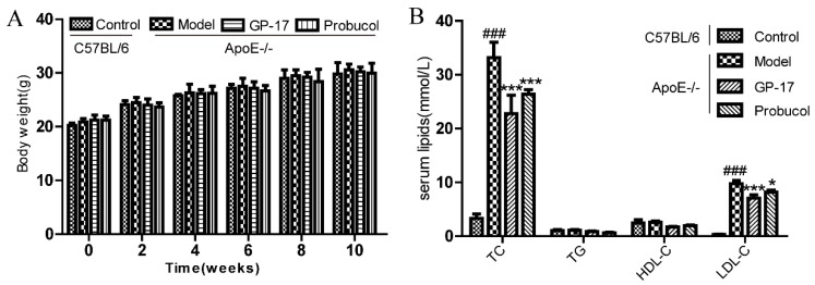
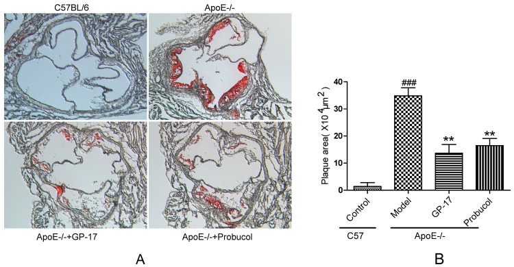
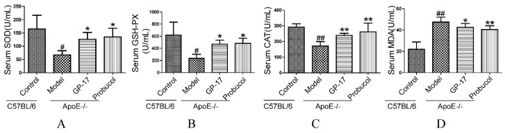
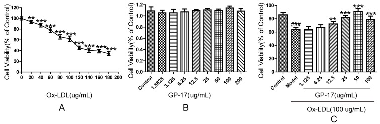
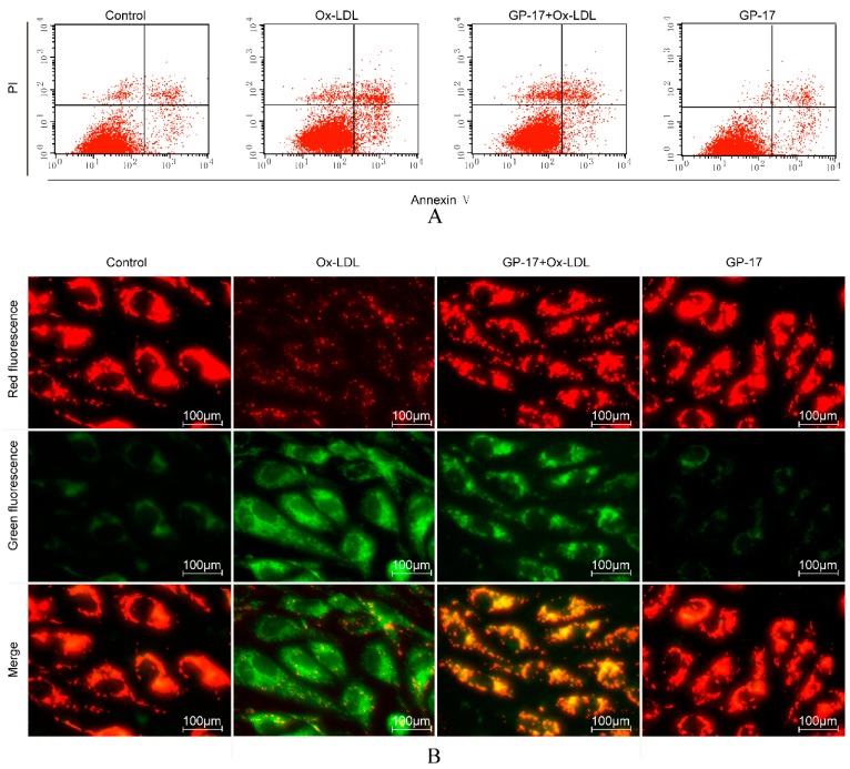
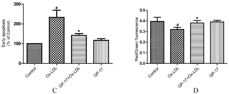
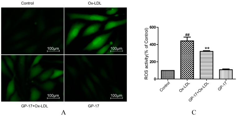
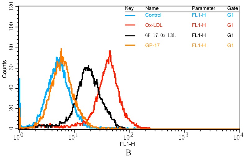
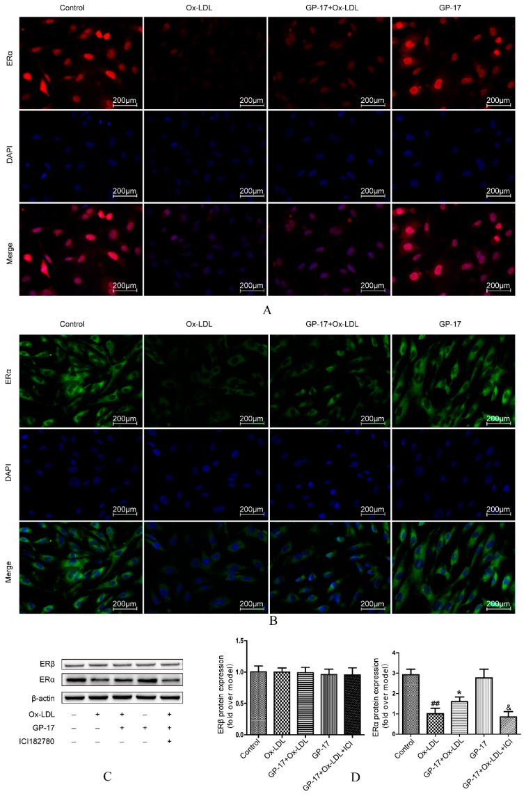
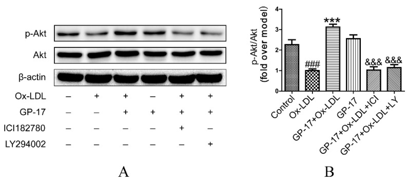
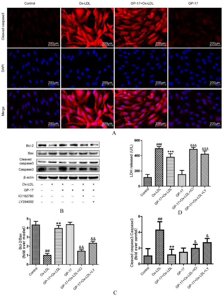
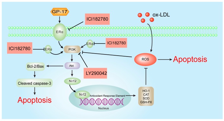
Phytoestrogens are estrogen-like compounds of plant origin. The pharmacological activities of phytoestrogens are predominantly due to their antioxidant, anti-inflammatory and lipid-lowering properties, which are mediated via the estrogen receptors (ERs): estrogen receptor alpha (ERα) and estrogen receptor beta (ERβ) and possibly G protein-coupled estrogen receptor 1 (GPER). Gypenoside XVII (GP-17) is a phytoestrogen that is widely used to prevent cardiovascular disease, including atherosclerosis, but the mechanism underlying these therapeutic effects is largely unclear. This study aimed to assess the anti-atherogenic effects of GP-17 and its mechanisms in vivo and in vitro. In vivo experiments showed that GP-17 significantly decreased blood lipid levels, increased the expression of antioxidant enzymes and decreased atherosclerotic lesion size in ApoE-/- mice. In vitro experiments showed that GP-17 significantly prevented oxidized low-density lipoprotein (Ox-LDL)-induced endothelial injury. The underlying protective mechanisms of GP-17 were mediated by restoring the normal redox state, up-regulating of the ratio of Bcl-2 to Bax and inhibiting the expression of cleaved caspase-3 in Ox-LDL-induced human umbilical vein endothelial cell (HUVEC) injury. Notably, we found that GP-17 treatment predominantly up-regulated the expression of ERα but not ERβ. However, similar to estrogen, the protective effect of GP-17 could be blocked by the ER antagonist ICI182780 and the phosphatidylinositol 3-kinase (PI3K) antagonist LY294002. Taken together, these results suggest that, due to its antioxidant properties, GP-17 could alleviate atherosclerosis via the ERα-mediated PI3K/Akt pathway.

Keywords: apoptosis; atherosclerosis; estrogen receptors; gypenoside XVII; oxidative damage.


Conflict of interest statement

The authors declare no conflict of interest.

Figures

Figure 1
Figure 1
The chemical structure of GP-17.
Figure 2
Figure 2
Effects of GP-17 on the body weights and serum lipids at 10 weeks in high fat diet-induced atherosclerotic ApoE−/− mice. (A) Changes in body weight after treatment; (B) The level of serum lipids. The values are expressed as the mean ± S.E.M. (n = 8 for each group). ### p < 0.001 vs. Control; * p < 0.05; *** p < 0.01 vs. Model. TC, total cholesterol; TG, triglyceride; HDL-C, high-density lipoprotein cholesterol; LDC-C, low-density lipoprotein cholesterol.
Figure 3
Figure 3
GP-17 effects on lipid deposition in the aortic sinus stained with oil red O. (A) Atherosclerotic lesion shown with oil red O staining; (B) Quantitative analysis of plaque areas in the aortic sinus by Image-Pro Plus software. Original magnification (A): ×200. The values are expressed as the mean ± S.E.M. (n = 8 for each group). ### p < 0.001 vs. Control; ** p < 0.01 vs. Model.
Figure 4
Figure 4
GP-17 increased the levels of SOD, GSH-Px and CAT and decreased MDA content in the serum. (A) Serum SOD level; (B) Serum GSH-Px level; (C) Serum CAT level; (D) Serum MDA level. The values are expressed as the mean ± S.E.M. (n = 8 for each group). # p < 0.05; ## p < 0.01 vs. Control; * p < 0.05; ** p < 0.01 vs. Model.
Figure 5
Figure 5
Cytoprotective effects of GP-17 on Ox-LDL-induced cytotoxicity in HUVECs. (A) HUVECs were treated with Ox-LDL at different concentrations for 24 h. Cell viability was measured by MTT assays; (B) Cell viability of HUVECs incubated with different concentrations of GP-17 for 12 h; (C) Incubation with GP-17 significantly lowered Ox-LDL-induced cell injury. Cell viability was measured by MTT assays. The values are expressed as the mean ± S.E.M. from three independent experiments. ### p < 0.001 vs. Control; ** p < 0.01; *** p < 0.001 vs. Model. MTT, 3-(4,5-dimethylthiazol-2yl)-2,5-diphenyltetrazolium bromide.
Figure 6
Figure 6
GP-17 protected against Ox-LDL-induced apoptosis in HUVECs. (A) Apoptosis in HUVECs was analyzed by flow cytometry; (B) ΔΨm was assessed by fluorescence microscopy; (C) Quantitative analysis of the percentages of early apoptotic cells; (D) Quantitative analysis of ΔΨm by plate reader. The values are expressed as the mean ± S.E.M. from three independent experiments. # p < 0.05 vs. Control; * p < 0.05 vs. Ox-LDL. PI, propidium iodide.
Figure 6
Figure 6
GP-17 protected against Ox-LDL-induced apoptosis in HUVECs. (A) Apoptosis in HUVECs was analyzed by flow cytometry; (B) ΔΨm was assessed by fluorescence microscopy; (C) Quantitative analysis of the percentages of early apoptotic cells; (D) Quantitative analysis of ΔΨm by plate reader. The values are expressed as the mean ± S.E.M. from three independent experiments. # p < 0.05 vs. Control; * p < 0.05 vs. Ox-LDL. PI, propidium iodide.
Figure 7
Figure 7
Effects of GP-17 on Ox-LDL-induced ROS production in HUVECs. (A) The ROS level was analyzed by fluorescence imaging; (B,C) Quantitative analysis of the fluorescence intensity of cells by flow cytometry. The values are expressed as the mean ± S.E.M. from three independent experiments. ## p < 0.01 vs. Control; ** p < 0.01 vs. Ox-LDL.
Figure 7
Figure 7
Effects of GP-17 on Ox-LDL-induced ROS production in HUVECs. (A) The ROS level was analyzed by fluorescence imaging; (B,C) Quantitative analysis of the fluorescence intensity of cells by flow cytometry. The values are expressed as the mean ± S.E.M. from three independent experiments. ## p < 0.01 vs. Control; ** p < 0.01 vs. Ox-LDL.
Figure 8
Figure 8
Effect of GP-17 on ERα in Ox-LDL-treated HUVECs. (A) Localization of ERα immunoreactivity was identified in the cytoplasm by immunofluorescence assays; (B) Localization of ERα immunoreactivity was identified in the plasma membrane by immunofluorescence assay; (C,D) The expressions of both ERα and ERβ were analyzed by western blots of Ox-LDL-induced HUVECs. The values are expressed as the mean ± S.E.M. from three independent experiments. ## p < 0.01 vs. Control; * p < 0.05 vs. Ox-LDL; & p < 0.05 vs. GP-17 + Ox-LD. DAPI, 4,6-diamidino-2-phenylindole.
Figure 9
Figure 9
Effect of GP-17 on PI3K/Akt phosphorylation via ERα in Ox-LDL treated HUVECs. (A,B) Phosphorylation of Akt mediated by GP-17 in Ox-LDL-induced HUVECs was abolished by pretreatment with ICI182780 or LY294002. The values are expressed as the mean ± S.E.M. from three independent experiments. # p < 0.05; ### p < 0.001 vs. Control; * p < 0.05; *** p < 0.001 vs. Ox-LDL; & p < 0.05; &&& p < 0.001 vs. GP-17 + Ox-LDL. p-Akt, phospho-protein kinase B.
Figure 10
Figure 10
GP-17 protected HUVECs against Ox-LDL-induced oxidative stress by increasing the antioxidant enzymes and antioxidant proteins. (A) The expression of HO-1 was analyzed by immunofluorescence assays; (B,C) The expressions of antioxidant proteins mediated by GP-17 in Ox-LDL-induced HUVECs were abolished by pretreatment with ICI182780 or LY294002; (D) the levels of SOD, GSH-Px and CAT activities and MDA production mediated by GP-17 in Ox-LDL-induced HUVEC injury were determined and abolished by pretreatment with ICI182780 or LY294002. The values are expressed as the mean ± S.E.M. from three independent experiments. ## p < 0.01; ### p < 0.001 vs. Control; * p < 0.05; ** p < 0.01; *** p < 0.001 vs. Ox-LDL; & p < 0.05; && p < 0.01; &&& p < 0.001 vs. GP-17 + Ox-LDL. HO-1, heme oxygenase 1; SOD, superoxide dismutase; GSH-Px, glutathione peroxidase; CAT, catalase; MDA, malondialdehyde.
Figure 11
Figure 11
GP-17 protected HUVECs against Ox-LDL-induced apoptosis by decreasing the ratio of Bax to Bcl-2 and controlling cleaved caspase-3 activation. (A) The expression of cleaved caspase-3 was analyzed by immunofluorescence assays; (B,C) The expression of apoptosis-inducing proteins mediated by GP-17 in Ox-LDL-induced HUVECs was abolished by pretreatment with ICI182780 or LY294002; (D) The protective effect of GP-17 against LDH release induced by Ox-LDL in HUVECs. The values are expressed as the mean ± S.E.M. from three independent experiments. ## p < 0.01; ### p < 0.001 vs. Control; ** p < 0.01; *** p < 0.001 vs. Ox-LDL; & p < 0.05; && p < 0.01; &&& p < 0.001 vs. GP-17 + Ox-LDL.
Figure 12
Figure 12
The molecular mechanism of the GP-17 anti-atherosclerotic effects. ROS induced by Ox-LDL result in endotheliocyte apoptosis and oxidative stress. GP-17 activates the ERα-mediated PI3K/Akt pathway, further resulting in Nrf2/HO-1 up-regulation and increasing the level of antioxidant enzymes. This process attenuates Ox-LDL-induced oxidative injury. Furthermore. GP-17 inhibited Ox-LDL-mediated HUVEC apoptosis by decreasing the ratio of Bax to Bcl-2 and controlling cleaved caspase-3 activation. This process inhibits Ox-LDL-induced endothelial cell apoptosis via the ERα-mediated PI3K/Akt pathway. T bars represented inhibition and arrows represented activation.

References

    1. Ross R. Atherosclerosis—An inflammatory disease. N. Engl. J. Med. 1999;340:115–126. doi: 10.1016/S0002-8703(99)70266-8. - DOI - PubMed
    1. Napoli C. Oxidation of LDL, atherogenesis, and apoptosis. Ann. N. Y. Acad. Sci. 2003;1010:698–709. doi: 10.1196/annals.1299.127. - DOI - PubMed
    1. Botham K.M., Wheeler-Jones C.P. Postprandial lipoproteins and the molecular regulation of vascular homeostasis. Prog. Lipid Res. 2013;52:446–464. doi: 10.1016/j.plipres.2013.06.001. - DOI - PubMed
    1. Gao P., Wang X.M., Qian D.H., Qin Z.X., Jin J., Xu Q., Yuan Q.Y., Li X.J., Si L.Y. Induction of oxidative stress by oxidized LDL via meprinalpha-activated epidermal growth factor receptor in macrophages. Cardiovasc. Res. 2013;97:533–543. doi: 10.1093/cvr/cvs369. - DOI - PubMed
    1. Dandapat A., Hu C., Sun L., Mehta J.L. Small concentrations of oxLDL induce capillary tube formation from endothelial cells via LOX-1-dependent redox-sensitive pathway. Arterioscler. Thromb. Vasc. Biol. 2007;27:2435–2442. doi: 10.1161/ATVBAHA.107.152272. - DOI - PubMed

MeSH terms