. 2017 Feb 9;7(1):13.
doi: 10.3390/biom7010013.

Next-Generation Sequencing-Based RiboMethSeq Protocol for Analysis of tRNA 2'-O-Methylation

Affiliations

Next-Generation Sequencing-Based RiboMethSeq Protocol for Analysis of tRNA 2'-O-Methylation

Virginie Marchand et al. Biomolecules. .

Abstract

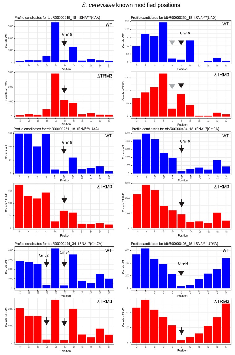
Analysis of RNA modifications by traditional physico-chemical approaches is labor intensive, requires substantial amounts of input material and only allows site-by-site measurements. The recent development of qualitative and quantitative approaches based on next-generation sequencing (NGS) opens new perspectives for the analysis of various cellular RNA species. The Illumina sequencing-based RiboMethSeq protocol was initially developed and successfully applied for mapping of ribosomal RNA (rRNA) 2'-O-methylations. This method also gives excellent results in the quantitative analysis of rRNA modifications in different species and under varying growth conditions. However, until now, RiboMethSeq was only employed for rRNA, and the whole sequencing and analysis pipeline was only adapted to this long and rather conserved RNA species. A deep understanding of RNA modification functions requires large and global analysis datasets for other important RNA species, namely for transfer RNAs (tRNAs), which are well known to contain a great variety of functionally-important modified residues. Here, we evaluated the application of the RiboMethSeq protocol for the analysis of tRNA 2'-O-methylation in Escherichia coli and in Saccharomyces cerevisiae. After a careful optimization of the bioinformatic pipeline, RiboMethSeq proved to be suitable for relative quantification of methylation rates for known modified positions in different tRNA species.

Keywords: tRNA; TrmH; 2′‐O‐methylation; RiboMethSeq; TRM3; deleted strain; high‐throughput sequencing.


Conflict of interest statement

The authors declare no conflict of interest.

Figures

Figure 1
Figure 1
Known positions of tRNA 2′-O-methylations and corresponding enzymes in Eubacteria (E. coli/Bacillus subtilis), Archaea (data almost exclusively from H. volcanii) and Eukarya (S. cerevisiae and various vertebrates). tRNAs are represented in folded 3D structures. 2′-O-methylated nucleotides are shown as spheres, orange for Eubacteria, pink for Archaea. Light blue color corresponds to yeast modifications; additional vertebrate positions are shown in dark blue. Names of homologous human proteins are in italics.
Figure 2
Figure 2
Fragmentation profiles for E. coli and S. cerevisiae total tRNA fractions. Alkaline hydrolysis was performed for 6–12 min (as indicated on the traces). Fragments size was analyzed by capillary electrophoresis on a Bioanalyzer 2100 (Agilent, Santa Clara, CA, USA) Small RNA Chip. FU: fluorescence unit.
Figure 3
Figure 3
Optimized treatment pipeline for tRNA 2′-O-methylation analysis by high-throughput sequencing. SAM: sequence alignment/map.
Figure 4
Figure 4
Proportion of non-uniquely mapped reads with initial and collapsed (optimized) transfer DNA (tDNA) datasets for each tRNA species. For both datasets, the number of uniquely and non-uniquely mapped reads was calculated for each tRNA species and plotted as stacked bars.
Figure 5
Figure 5
Representative cleavage profiles for six E. coli tRNA species for wild-type (WT) and ΔTrmH strains. The cumulated number of the 5′- and 3′-end extremities at every position is traced as vertical bars. All profiles are centered for the known 2′-O-methylated position shown by the black arrow; false-positive alkali-resistant positions are indicated by gray arrows.
Figure 6
Figure 6
Representative cleavage profiles for six S. cerevisiae tRNA species for WT and ΔTRM3 strains. The cumulated number of the 5′- and 3′-end extremities at every position is traced as vertical bars. All profiles are centered for the known 2′-O-methylated position shown by the black arrow; false-positive alkali-resistant positions are indicated by gray arrows.

References

    1. Cheng X., Roberts R.J. AdoMet-dependent methylation, DNA methyltransferases and base flipping. Nucleic Acids Res. 2001;29:3784–3795. doi: 10.1093/nar/29.18.3784. - DOI - PMC - PubMed
    1. Boschi-Muller S., Motorin Y. Chemistry enters nucleic acids biology: Enzymatic mechanisms of RNA modification. Biochem. Biokhimiia. 2013;78:1392–1404. doi: 10.1134/S0006297913130026. - DOI - PubMed
    1. Motorin Y., Seidu-Larry S., Helm M. DNA and RNA Pyrimidine Nucleobase Alkylation at the Carbon-5 Position. Adv. Exp. Med. Biol. 2016;945:19–33. - PubMed
    1. Rana A.K., Ankri S. Reviving the RNA World: An Insight into the Appearance of RNA Methyltransferases. Front. Genet. 2016;7:99. doi: 10.3389/fgene.2016.00099. - DOI - PMC - PubMed
    1. Towns W.L., Begley T.J. Transfer RNA methytransferases and their corresponding modifications in budding yeast and humans: Activities, predications, and potential roles in human health. DNA Cell Biol. 2012;31:434–454. doi: 10.1089/dna.2011.1437. - DOI - PMC - PubMed

MeSH terms

LinkOut - more resources