. 2017 Feb 16;14(1):33.
doi: 10.1186/s12985-017-0704-x.

Evaluation of the contribution of the transmembrane region to the ectodomain conformation of the human immunodeficiency virus (HIV-1) envelope glycoprotein

Affiliations

Evaluation of the contribution of the transmembrane region to the ectodomain conformation of the human immunodeficiency virus (HIV-1) envelope glycoprotein

Hanh T Nguyen et al. Virol J. .

Abstract

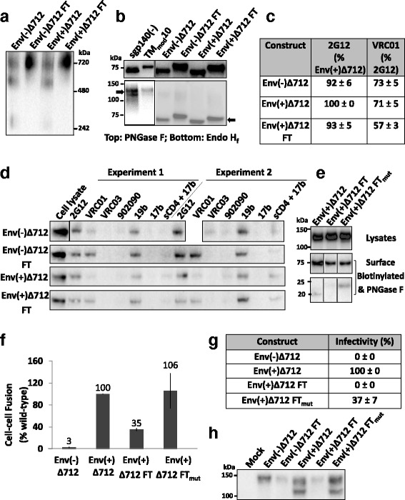
Background: The human immunodeficiency virus (HIV-1) envelope glycoprotein (Env), a Type 1 transmembrane protein, assembles into a trimeric spike complex that mediates virus entry into host cells. The high potential energy of the metastable, unliganded Env trimer is maintained by multiple non-covalent contacts among the gp120 exterior and gp41 transmembrane Env subunits. Structural studies suggest that the gp41 transmembrane region forms a left-handed coiled coil that contributes to the Env trimer interprotomer contacts. Here we evaluate the contribution of the gp41 transmembrane region to the folding and stability of Env trimers.

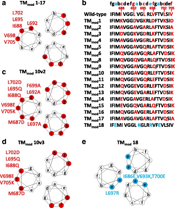
Methods: Multiple polar/charged amino acid residues, which hypothetically disrupt the stop-transfer signal, were introduced in the proposed lipid-interactive face of the transmembrane coiled coil, allowing release of soluble cleavage-negative Envs containing the modified transmembrane region (TMmod). We also examined effects of cleavage, the cytoplasmic tail and a C-terminal fibritin trimerization (FT) motif on oligomerization, antigenicity and functionality of soluble and membrane-bound Envs.

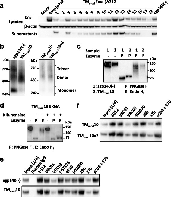
Results: The introduction of polar/charged amino acids into the transmembrane region resulted in the secretion of soluble Envs from the cell. However, these TMmod Envs primarily formed dimers. By contrast, control cleavage-negative sgp140 Envs lacking the transmembrane region formed soluble trimers, dimers and monomers. TMmod and sgp140 trimers were stabilized by the addition of a C-terminal FT sequence, but still exhibited carbohydrate and antigenic signatures of a flexible ectodomain structure. On the other hand, detergent-solubilized cleaved and uncleaved Envs isolated from the membranes of expressing cells exhibited "tighter" ectodomain structures, based on carbohydrate modifications. These trimers were found to be unstable in detergent solutions, but could be stabilized by the addition of a C-terminal FT moiety. The C-terminal FT domain decreased Env cleavage and syncytium-forming ability by approximately three-fold; alteration of the FT trimerization interface restored Env cleavage and syncytium formation to near-wild-type levels.

Conclusion: The modified transmembrane region was not conducive to trimerization of soluble Envs. However, for HIV-1 Env ectodomains that are minimally modified, membrane-anchored Envs exhibit the most native structures and can be stabilized by appropriately positioned FT domains.

Keywords: Ectodomain; Fibritin –Trimer; HIV-1 Env; Stabilize; Transmembrane region; gp41.


Figures

Fig. 1
Fig. 1
Location of changes in the transmembrane region of HIV-1JR-FL Envs. a The projection of the helical coiled coil in the HIV-1 Env transmembrane region, based on the NMR structure of a peptide embedded in bicelles [29], is shown. Six hydrophobic residues (I688, L692, L695, V698, L702 and V705) highlighted in red are predicted to be located on the lipid-interacting surface of the membrane-spanning coiled coil. b In the TMmod1-17 constructs, the six hydrophobic residues on the putative lipid-interacting surface in a were changed to combinations of alanine, glutamine or charged amino acid residues. c The TMmod10v2 construct is a TMmod10 variant with three additional changes (M687D, L697A and F699A) introduced to modify all of the predicted external positions b, c, e, f, g on the helical coiled coil. d The TMmod10v3 construct is identical to TMmod10v2 except that the residues in the e and g positions of the coiled coil (L692, L697 and F699) are wild-type in sequence. e The TMmod18 glycoprotein control contains four changes in the transmembrane region, three of which (I686E, V693K and T700E) are predicted to be located in the interior of the coiled coil (at the a position of the heptad repeat sequence)
Fig. 2
Fig. 2
Characterization of TMmod mutant Envs. a Cellular lysates and supernatants from 293T cells that were mock-transfected or transfected with TMmod Env DNAs were Western blotted. Western blots shown in this figure used goat anti-gp120 antibody or a mouse anti-β-actin control. The TMmod Envs with at least two new charged residues in the transmembrane region were secreted. The control TMmod18 Env was inefficiently expressed and not secreted. The sgp140(−) Env lacks the transmembrane region. b The secreted Envs were analyzed by Blue Native PAGE. The sgp140(−) glycoprotein migrated as a heterogeneous mixture of monomers, dimers and trimers. The representative TMmod10 protein migrated predominantly as a dimer. The TMmod10v2 glycoprotein was also largely dimeric. Note that HIV-1 Envs migrate more slowly than expected in Blue Native gels. c Transfected cell supernatants containing sgp140(−) and TMmod10 Envs were either mock-treated or treated with PNGase F (which removes all N-linked glycans) or Endo Hf (which removes only high-mannose glycans). The Western blot shows that both sgp140(−) and TMmod10 Envs resist Endo Hf treatment, which indicates that they contain mostly complex carbohydrates. d Transfected cells expressing the E168K + N188A (EKNA) variant of TMmod10, which allows HIV-1JR-FL Envs to be recognized by the PG9 and PG16 neutralizing antibodies [–99], were incubated with 50 mM kifunensine (a mannosidase I inhibitor). Cell supernatants were collected, deglycosylated with PNGase F or Endo Hf, and Western blotted. Addition of kifunensine converted TMmod10 glycosylation from mostly complex glycans to high-mannose glycans. e, f Cell supernatants containing the indicated soluble glycoproteins were precipitated with the indicated antibodies, and the precipitates were Western blotted with goat anti-gp120 antibody. The three soluble Envs exhibit a similar pattern of antigenicity. One-fourth volume of the supernatant used for immunoprecipitation was analyzed in the input lane. Data are representative of those obtained in at least two independent experiments
Fig. 3
Fig. 3
Effects of cytoplasmic tail and cleavage site modifications on TMmod10 Env. a The TMmod10 EKNA variant has the E168K + N188A changes that allow the HIV-1JR-FL Env to be recognized by the PG9 and PG16 antibodies [–99]. The full-length TMmod10 EKNA Env or the TMmod10 EKNA variants with a deleted (Δ712) or truncated (Δ753 and Δ808) cytoplasmic tail were expressed in 293T cells. The cell lysates and supernatants were analyzed by SDS-PAGE, and the cell supernatants by Blue Native PAGE. The gels shown in this figure were Western blotted with a polyclonal goat anti-gp120 antibody. Addition of the cytoplasmic tail did not prevent TMmod10 EKNA expression in cells, but only the TMmod10Δ753 EKNA glycoprotein with the shortest tail was secreted. The secreted TMmod10Δ753 Env was mainly dimeric based on the Blue Native gel. b The TMmodΔ712 and TMmodΔ753 EKNA Envs precipitated from the supernatants of expressing cells by the indicated antibodies are shown. The antigenic profiles of these two Envs are similar. c TMmod10 EKNA variants with modifications of the cleavage site, including a wild-type cleavage site (+) or a flexible linker ((GGS)4) replacing the cleavage site (modCS), were analyzed as in a. Note that the TMmod10 (+) EKNA Env is partially cleaved, as indicated by the presence of a gp120 band on SDS-PAGE. d Precipitation of the TMmod10 and TMmod10 EKNA variants by the indicated antibodies is shown. Data are representative of those obtained in at least two independent experiments
Fig. 4
Fig. 4
Effect of a fibritin trimerization motif on the TMmod10 Env. a Cell lysates and supernatants from 293T cells expressing the EKNA variants of sgp140(−) or TMmod10, or these Envs with a C-terminal fibritin trimerization domain (sgp140(−) FT and TMmod10 FT, respectively), were analyzed on gels and Western blotted. A polyclonal goat anti-gp120 antibody was used to detect the Envs on the Western blots shown in this figure. Addition of the fibritin domain to the C-terminus of TMmod10 did not prevent Env expression but significantly diminished release of the TMmod10 FT glycoprotein from the cells. b The indicated soluble Envs were subjected to digestion with PNGase F or Endo Hf and Western blotted. c Antibody precipitation of the EKNA variants of sgp140(−), sgp140(−) FT, TMmod10 and TMmod10 FT Envs secreted into the medium of expressing cells is shown. All EKNA Env constructs contain the E168K + N188A changes that restore the PG9/PG16 epitopes [–99]. Data are representative of those obtained in duplicate or two independent experiments
Fig. 5
Fig. 5
Effect of the C-terminal fibritin trimerization motif on membrane-anchored Envs. a Envs purified from transfected 293T cell membranes were analyzed by Blue Native PAGE. The Env(−)Δ712 FT and Env(+)Δ712 FT glycoproteins migrate at a size expected for trimers. b The purified soluble or membrane Envs were treated with PNGase F (top panel) or Endo Hf (bottom panel). The arrows indicate Envs after Endo Hf digestion. Compared to sgp140(−) and TMmod10 Envs, which are relatively resistant to Endo Hf digestion, purified membrane Envs are Endo Hf-sensitive, and thus rich in high-mannose carbohydrates. c Flow cytometry was used to study Env surface expression and recognition by the 2G12 glycan-dependent antibody and the VRC01 CD4-binding site antibody. d To immunoprecipitate cell-surface Env, 293T cells transiently expressing the indicated Envs were incubated with antibodies, washed and lysed. Cell lysates were incubated with Protein A-Sepharose beads. Precipitates were Western blotted with a goat anti-gp120 antibody. e To assess cell-surface Env processing, cells expressing the indicated Envs were biotinylated as described in Methods. The cell lysates (upper panel) or deglycosylated cell-surface Envs (lower panels) were Western blotted with the 4E10 anti-gp41 antibody. After deglycosylation, the uncleaved Env is 75 kD, and the cleaved transmembrane Envs are 20–23 kD. f An α-complementation assay was used to measure Env-mediated cell-cell fusion. The reduced cell-cell fusion activity of Env(+)Δ712 FT was restored by the disruption of fibritin trimerization in the Env(+)Δ712 FTmut glycoprotein. g The infectivity of recombinant luciferase-expressing HIV-1 with the indicated Envs was measured on Cf2Th-CD4/CCR5 target cells. The luciferase activity in the target cells was normalized to that observed for the Env(+)Δ712 glycoprotein. h Virions pseudotyped with the indicated Env proteins were purified through a 20% sucrose cushion, denatured and Western blotted. Data in this figure are representative of, or averaged from, those obtained in at least two independent experiments. Error bars are standard deviations

Similar articles

Cited by

References

    1. Wyatt R, Sodroski J. The HIV-1 envelope glycoproteins: fusogens, antigens, and immunogens. Science. 1998;280:1884–1888. doi: 10.1126/science.280.5371.1884. - DOI - PubMed
    1. Allan JS, Coligan JE, Barin F, McLane MF, Sodroski JG, Rosen CA, Haseltine WA, Lee TH, Essex M. Major glycoprotein antigens that induce antibodies in AIDS patients are encoded by HTLV-III. Science. 1985;228:1091–1094. doi: 10.1126/science.2986290. - DOI - PubMed
    1. Robey WG, Safai B, Oroszlan S, Arthur LO, Gonda MA, Gallo RC, Fischinger PJ. Characterization of envelope and core structural gene products of HTLV-III with sera from AIDS patients. Science. 1985;228:593–595. doi: 10.1126/science.2984774. - DOI - PubMed
    1. Pal R, Hoke GM, Sarngadharan MG. Role of oligosaccharides in the processing and maturation of envelope glycoproteins of human immunodeficiency virus type 1. Proc Natl Acad Sci U S A. 1989;86:3384–3388. doi: 10.1073/pnas.86.9.3384. - DOI - PMC - PubMed
    1. Earl PL, Moss B, Doms RW. Folding, interaction with GRP78-BiP, assembly, and transport of the human immunodeficiency virus type 1 envelope protein. J Virol. 1991;65:2047–2055. - PMC - PubMed

Publication types

MeSH terms

Substances

LinkOut - more resources