. 2017 Apr:124:169-179.
doi: 10.1016/j.biomaterials.2017.02.002. Epub 2017 Feb 4.

Exploring deformable particles in vascular-targeted drug delivery: Softer is only sometimes better

Affiliations

Exploring deformable particles in vascular-targeted drug delivery: Softer is only sometimes better

Margaret B Fish et al. Biomaterials. 2017 Apr.

Abstract

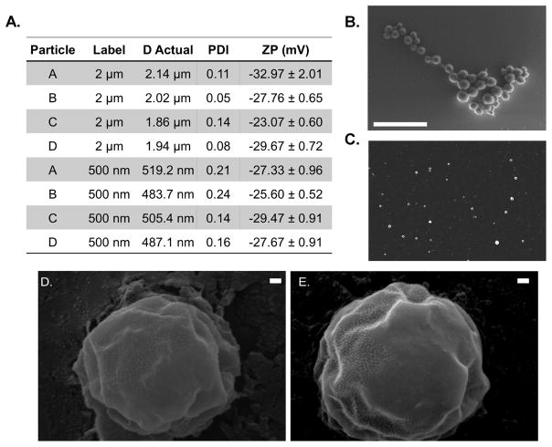
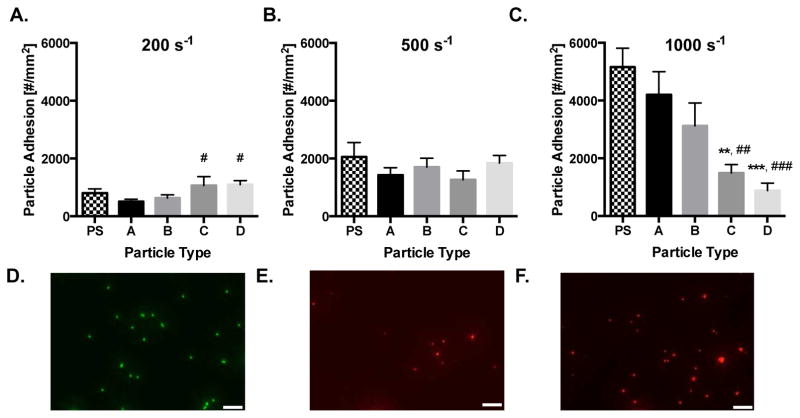
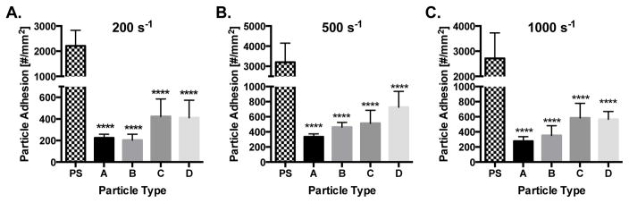
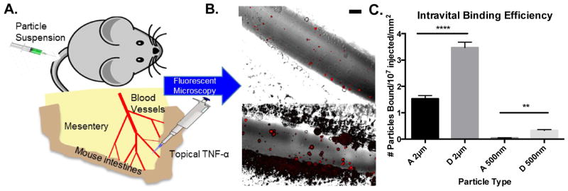
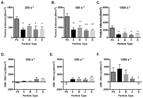
The ability of vascular-targeted drug carriers (VTCs) to localize and bind to a targeted, diseased endothelium determines their overall clinical utility. Here, we investigate how particle modulus and size determine adhesion of VTCs to the vascular wall under physiological blood flow conditions. In general, deformable microparticles (MPs) outperformed nanoparticles (NPs) in all experimental conditions tested. Our results indicate that MP modulus enhances particle adhesion in a shear-dependent manner. In low shear human blood flow profiles in vitro, low modulus particles showed favorable adhesion, while at high shear, rigid particles showed superior adhesion. This was confirmed in vivo by studying particle adhesion under venous shear profiles in a mouse model of mesenteric inflammation, where MP adhesion was 127% greater (p < 0.0001) for low modulus particles compared to more rigid ones. Mechanistically, we establish that particle collisions with leukocytes drive these trends, rather than differences in particle deformation, localization, or detachment. Overall, this work demonstrates the importance of VTC modulus as a design parameter for enhanced VTC interaction with vascular walls, and thus, contributes important knowledge for development of successful clinical theranostics with applications for many diseases.

Keywords: Deformability; Hemodynamics; Hydrogel; Modulus; Shear force; Vascular-targeted carrier.


Conflict of interest statement

Conflict of interest statement: The authors have declared that no conflict of interest exists.

Figures

Figure 1
Figure 1. Hydrogel material properties
(A) Designation of chemical moieties for synthetic scheme. (B) Synthetic scheme for lithium phenyl-2,4,6-trimethylbenzoylphosphinate photoinitiated polymerization of (poly)ethylene glycol diacrylate (PEGDA) and 2-carboxylethyl acrylate (CEA) as described in more detail in the SI. (C) In situ rheometry of particle conditions B, C, and D. Condition A too rigid to be tested in situ. (D) Swollen shear moduli of particle conditions A–D, statistics displayed represent comparison to A. (E) Equilibrium swelling ratios of particle conditions A–D, where (*s) indicate significance within hydrogel types to A and (#s) indicate difference between water and plasma. (F) Synthesis compositions and calculated bulk material properties of hydrogels. Statistical analyses were performed using one- and two-way ANOVA with Fisher’s LSD test, where (*) indicates p<0.05, (**) indicates p<0.01, and (***) indicates p<0.001, (****) indicates p<0.0–01 and (####) indicates p<0.0001. Error bars represent standard error.
Figure 2
Figure 2. Hydrogel particle properties
(A) Diameter, PDI and zeta potential measurements for fabricated hydrogel particles +/− standard deviation. Representative SEM Image of dried (B) 2 μm and (C) 500 nm hydrogel condition A particles, scale bars are 5 μm. Representative scanning electron micrographs at 50,000 X magnification of (D) Hydrogel particle type A and (E) D. Scale bars are 100 nm in length.
Figure 3
Figure 3. Particle adhesion to inflamed HUVEC monolayer as a function of particle modulus
Quantified adhesion of 2 μm hydrogel particles at a wall shear rate of (A) 200 s−1, (B) 500 s−1, and (C) 1,000 s−1 by modulus after 5 mins of laminar blood flow over an IL-1β activated HUVEC monolayer. N=3–6 human blood donors per particle condition. Statistical analysis of adherent density was performed using one-way ANOVA with Fisher’s LSD test between all particle adhesion conditions. (*) Represent comparison to PS and (#) represents comparison to particle type A. (*) indicates p<0.05, (**) indicates p<0.01, and (***) indicates p<0.001. Error bars represent standard error. Representative fluorescent images of particles bound to IL-1β activated HUVEC under a WSR of 200 s−1 in vitro for 2 μm (D) PS, (E) Hydrogel A, and (F) Hydrogel D. Scale bars are 20 μm.
Figure 4
Figure 4. 500 nm particle adhesion to inflamed HUVEC monolayer as a function of particle modulus
Quantified adhesion of hydrogel particles at a wall shear rate of (A) 200 s−1, (B) 500 s−1, and (C) 1,000 s−1 by modulus after 5 mins of laminar blood flow over an IL-1β activated HUVEC monolayer at 1 × 10⁸ particles/mL. N=3–6 human blood donors per particle condition. Statistical analysis of adherent density was performed using one-way ANOVA with Fisher’s LSD test between all particle adhesion conditions. (*) Represent comparison to PS where (*) indicates p<0.05, (**) indicates p<0.01, and (***) indicates p<0.001. There were no significant differences amongst hydrogel particle types A–D within any wall shear rate. Error bars represent standard error.
Figure 5
Figure 5
Particle adhesion to inflamed mesentery endothelium as a function of modulus and size. (A) Schematic of mouse surgical technique for intravital imaging, as described in the methods. (B) Representative fluorescence images of particle adhesion to inflamed mesentery, images correspond particles A-2μm and D-2μm (top to bottom). Particle fluorescence shown in red, overlaid on the bright field image. Scale bar is 50 μm. (C) Quantified adhesion efficiency of hydrogel particle conditions A-2μm, D-2μm, A-500nm, and D-500nm, scaled by vessel area, n = 4 mice per group. Statistical analysis was performed using one-way ANOVA with Fisher’s LSD test within particle sizes. (**) indicates p<0.01 and (****) indicates p<0.0001. Error bars represent standard error.
Figure 6
Figure 6. Hydrogel particle behavior under shear forces
(A) Strain response of bulk hydrogels A–D at controlled, applied shear stress. Arrows represent corresponding wall shear rates. (B) Deformability parameter (Δ) determined by FEA for particles A–D under a range of shear forces. Inset shows applied shear force directions and representative particle deformation of particle D under the largest shear (circled) with an amplification factor of 2 to visualize deformation. (C) Localization of fluorescent hydrogel particles from human whole blood flow to the chamber wall. N = 3 human blood donors per particle condition. Statistical analysis was performed using two-way ANOVA with Fisher’s LSD test between groups, resulting in non-significant differences at all shear rates. Error bars represent the standard error. (D) Representative rate of attachment and detachment to inflamed HUVECs at wall shear rates of 1000 s−1. The first 300 seconds represent perfusion of whole human blood and hydrogel particles, while the remaining time represents perfusion of buffer without particles.
Figure 7
Figure 7. Particle adhesion in WBC depleted, RBC+plasma medium to inflamed HUVEC monolayer
Quantified adhesion of 2 μm hydrogel particles in RBC+plasma at a wall shear rate of (A) 200 s−1, (B) 500 s−1, and (C) 1,000 s−1 by modulus after 5 mins of laminar flow over an IL-1β activated HUVEC monolayer. N=3–6 human blood donors per particle condition in WBC depleted RBC+plasma medium. Particle adhesion in whole blood minus adhesion in RBC+plasma is quantified for (D) 200 s−1, (E) 500 s−1, (F) 1,000 s−1 by modulus. Positive values signify WBCs enhance particle binding in shear flow. Statistical analysis of adherent density was performed using one-way ANOVA with Fisher’s LSD test between all particle adhesion conditions. (*) Represent comparison to PS and (#) represents comparison to particle type A. (*) indicates p<0.05, (**) indicates p<0.01, and (***) indicates p<0.001. Error bars represent standard error.

References

    1. Fish MB, Thompson AJ, Fromen CA, Eniola-Adefeso O. Emergence and Utility of Non-Spherical Particles in Biomedicine. Ind Eng Chem Res. 2015;54:4043–4059. - PMC - PubMed
    1. Albanese A, Tang PS, Chan WCW. The Effect of Nanoparticle Size, Shape, and Surface Chemistry on Biological Systems. Annu Rev Biomed Eng. 2012;14:1–16. - PubMed
    1. Torchilin VP. Multifunctional Nanocarriers. Adv Drug Deliv Rev. 2012;64:302–315. - PubMed
    1. Brannon-Peppas L, Blanchette JO. Nanoparticle and Targeted Systems for Cancer Therapy. Adv Drug Deliv Rev. 2012;6:206–212. - PubMed
    1. Sen Gupta A. Role of Particle Size, Shape, and Stiffness in Design of Intravascular Drug Delivery Systems: Insights from Computations, Experiments, and Nature. WIREs. 2016;8:255–270. - PubMed

Publication types

MeSH terms