. 2017 Apr 25;131(8):699-713.
doi: 10.1042/CS20170047. Epub 2017 Feb 16.

Human DPSCs fabricate vascularized woven bone tissue: a new tool in bone tissue engineering

Affiliations

Human DPSCs fabricate vascularized woven bone tissue: a new tool in bone tissue engineering

Francesca Paino et al. Clin Sci (Lond). .

Abstract

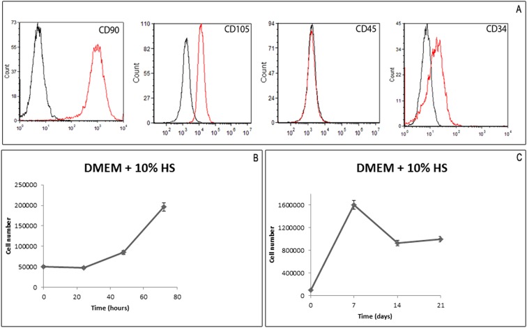
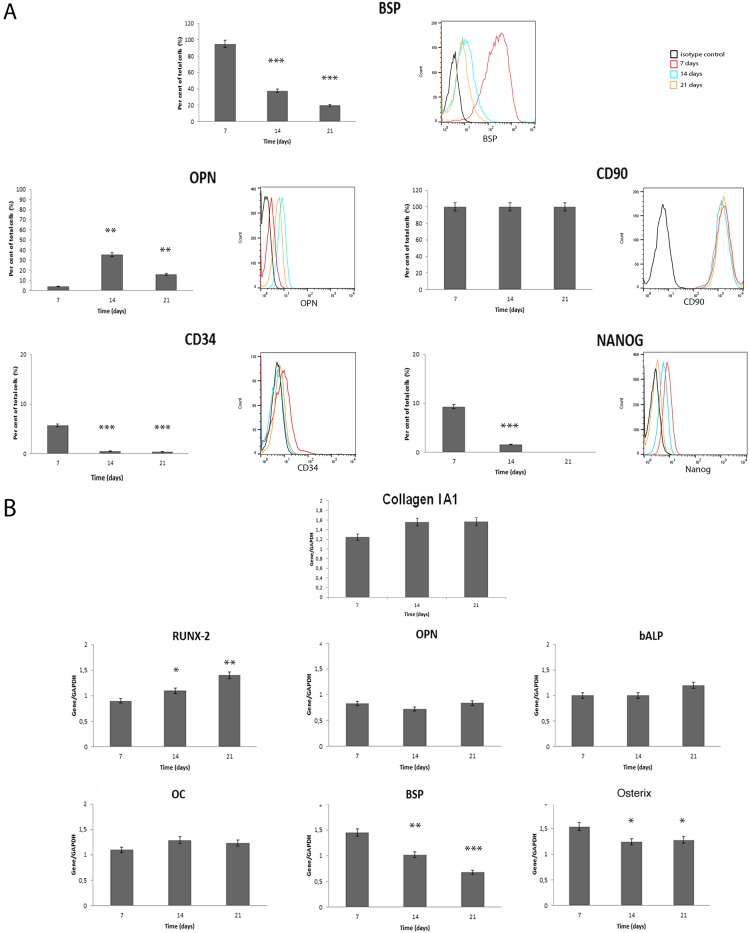
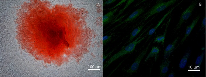
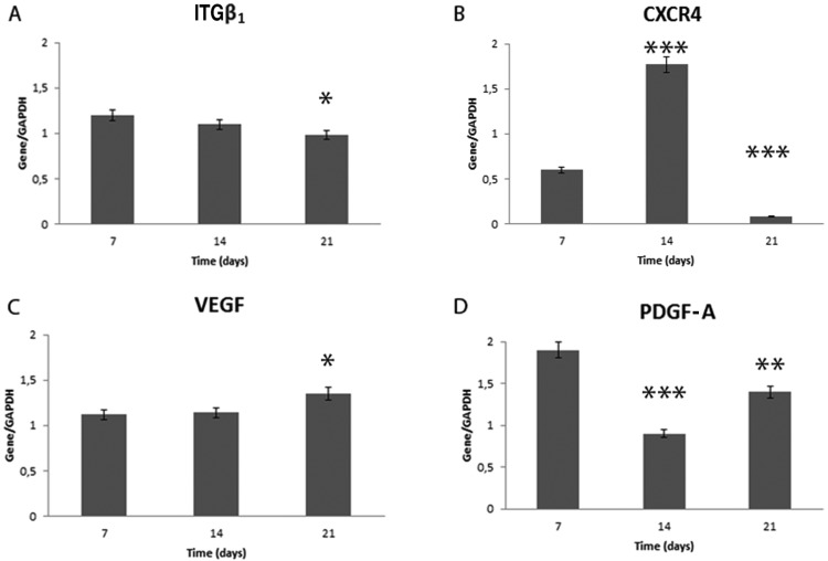
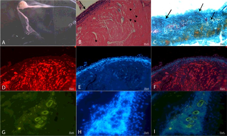
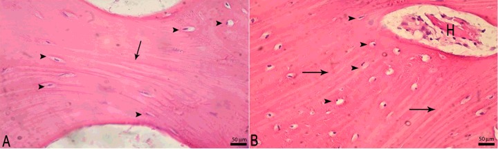
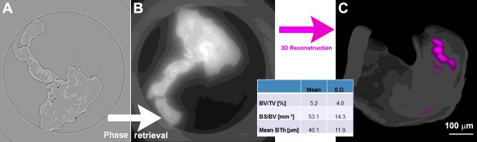
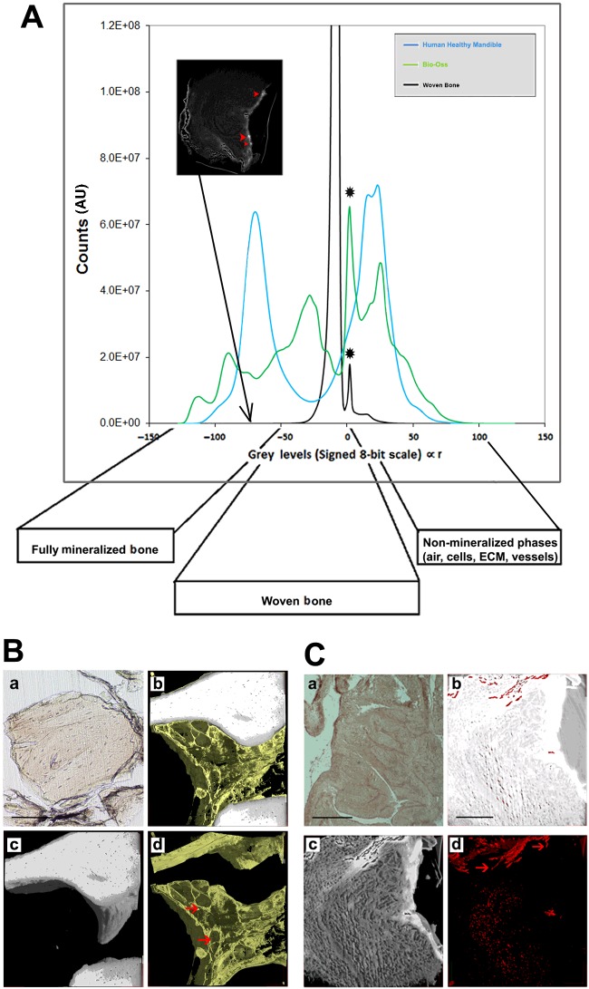
Human dental pulp stem cells (hDPSCs) are mesenchymal stem cells that have been successfully used in human bone tissue engineering. To establish whether these cells can lead to a bone tissue ready to be grafted, we checked DPSCs for their osteogenic and angiogenic differentiation capabilities with the specific aim of obtaining a new tool for bone transplantation. Therefore, hDPSCs were specifically selected from the stromal-vascular dental pulp fraction, using appropriate markers, and cultured. Growth curves, expression of bone-related markers, calcification and angiogenesis as well as an in vivo transplantation assay were performed. We found that hDPSCs proliferate, differentiate into osteoblasts and express high levels of angiogenic genes, such as vascular endothelial growth factor and platelet-derived growth factor A. Human DPSCs, after 40 days of culture, give rise to a 3D structure resembling a woven fibrous bone. These woven bone (WB) samples were analysed using classic histology and synchrotron-based, X-ray phase-contrast microtomography and holotomography. WB showed histological and attractive physical qualities of bone with few areas of mineralization and neovessels. Such WB, when transplanted into rats, was remodelled into vascularized bone tissue. Taken together, our data lead to the assumption that WB samples, fabricated by DPSCs, constitute a noteworthy tool and do not need the use of scaffolds, and therefore they are ready for customized regeneration.

Keywords: bone differentiation; bone regeneration; bone tissue engineering; hDPSCs; holotomography; human Dental Pulp Stem Cells; human serum; phc-microCT; woven bon; woven bone.


Figures

Figure 1
Figure 1. Characterization and growth of DPSCs
(A) Characterization of DPSCs at the first passage of culture: cytometric analysis of CD90, CD105, CD45 and CD34 markers in DPSCs. Histograms represent the number of cells (y-axis) and fluorescence intensity (x-axis) relative to unstained control cells (black histogram), and cells marked with specific antibodies against surface proteins (red histogram). (B) Growth curve of DPSCs: the average DT is 49±2 h. (C) Growth performance was studied for up to 21 days.
Figure 2
Figure 2. Cytometric analysis and gene expression in DPSCs
(A) Cytometric analysis and (B) gene expression for osteogenic markers, and stemness markers (L-N) in DPSCs. *P<0.001, **P<0.0005, ***P<0.0001 compared with the cell line at day 7.
Figure 3
Figure 3. Alizarin Red analysis of DPSCs
(A) Representative images showing Alizarin Red staining. Scale bar=100 μm. (B) Immunofluorescence analysis for OC. Scale bar=10 μm.
Figure 4
Figure 4. Reverse transcription PCR for genes involved in chemotaxis, cell adhesion and angiogenesis
In (A) and (B) are shown, respectively, expression of ITGβ1 and CXCR4 mRNA in DPSCs. In (C) and (D) VEGF and PDGF-A mRNA levels are shown in DPSCs. All results were normalized to GAPDH expression. *P<0.001, **P<0.0005, ***P<0.0001 compared with the cell line at day 7.
Figure 5
Figure 5. In vivo subcutaneous transplantation of WB into rats
(A) In the flask, a single WB sample measuring 1×1 cm is shown; (B) H&E stain showing in vivo bone tissue formation, in which osteocytes (arrowheads) and periosteum (arrows) can be observed; (C) Mallory's Trichrome stain showing in vivo vessel neoformation (arrows); (D) class I HLA positivity on in vivo bone tissue sample; (E) Hoechst stain of nuclei; (F) class I HLA positivity merge on in vivo bone tissue sample. (G) CD34 positivity on in vivo bone tissue sample; (H) Hoechst stain of nuclei; (I) CD34 positivity merges on in vivo bone tissue sample. Scale bars (C, G, H, I)=10 and (B, D, E, F)=50 μm.
Figure 6
Figure 6. In vivo transplantation of WB in a mandibular vertical defect
(A) H&E stain showing in vivo lamellar bone tissue formation with osteocytes (arrowheads) and lamellae (arrow); and (B) haversian canal formation (H) surrounded by lamellae and osteocytes in their lacunae (arrowheads). Scale bars=50 μm.
Figure 7
Figure 7. Phase-contrast microCT analysis of WB
(A) Two-dimensional slice without phase-retrieval processing: the edge-enhancement signal prevents a reliable discrimination and quantification of the two phases (WB and newly mineralized bone). (B) The same 2D slice as in (A) but after phase-retrieval processing; and (C) 3D reconstruction of the 2D slices previously processed by phase retrieval: the woven structure is shown in translucent white, whereas the newly formed mineralized bone is depicted in magenta; (bottom inset) morphometric analysis of the 3D volumes.
Figure 8
Figure 8. Histograms and 3D images of WB, Bio-Oss control and a human mandibular healthy site
(A) Grey-value distribution histograms of the samples obtained from WB, Bio-Oss control and a human mandibular healthy site. Peaks indicated with * are faked and due to mismatches of the samples in the field of view. (Top inset): 2D HT slice indicating few clusters of fully mineralized bone. (B) Three-dimensional images of Bio-Oss: (panel a) histological section with H&E staining, as a reference; (panels b–d) subvolume of the 3D HT reconstruction. To improve visualization, in each 3D image all phases were virtually deleted, except for: (b) bone and ECM, (c) bone and (d) ECM. Scale bar=250 μm. (C) Three-dimensional images of WB sample: (a) histological section with H&E staining; (panels b–d) subvolume of the 3D HT reconstruction. To improve visualization, in each 3D image all phases were virtually deleted, except for: (b) woven bone and vessels, (c) WB and (d) vessels. Red arrowheads indicate portions of vessels to distinguish them from possible artefacts. Scale bar=250 μm.

References

    1. Caplan A.I. (2007) Adult mesenchymal stem cells for tissue engineering versus regenerative medicine. J. Cell. Physiol. 213, 341–34710.1002/jcp.21200 - DOI - PubMed
    1. Ferro F., Spelat R., Beltrami A.P., Cesselli D. and Curcio F. (2012) Isolation and characterization of human dental pulp derived stem cells by using media containing low human serum percentage as clinical grade substitutes for bovine serum. PLoS One 7, e48945.10.1371/journal.pone.0048945 - DOI - PMC - PubMed
    1. Laino G., d'Aquino R., Graziano A., Lanza V., Carinci F., Naro F.. et al. (2005) A new population of human adult dental pulp stem cells: a useful source of living autologous fibrous bone tissue (LAB). J. Bone Miner. Res. 20, 1394–140210.1359/JBMR.050325 - DOI - PubMed
    1. d'Aquino R., Graziano A., Sampaolesi M., Laino G., Pirozzi G., De Rosa A.. et al. (2007) Human postnatal dental pulp cells codifferentiate into osteoblasts and endotheliocytes: a pivotal synergy leading to adult bone tissue formation. Cell Death Differ 14, 1162–117110.1038/sj.cdd.4402121 - DOI - PubMed
    1. Lizier N.F., Kerkis A., Gomes C.M., Hebling J., Oliveira C.F., Caplan A.I.. et al. (2012) Scaling-up of dental pulp stem cells isolated from multiple niches. PLoS ONE 7, e39885.10.1371/journal.pone.0039885 - DOI - PMC - PubMed

Publication types

MeSH terms