. 2017 Apr 14;292(15):6225-6239.
doi: 10.1074/jbc.M116.760314. Epub 2017 Feb 16.

Pathogenic mutations in retinitis pigmentosa 2 predominantly result in loss of RP2 protein stability in humans and zebrafish

Affiliations

Pathogenic mutations in retinitis pigmentosa 2 predominantly result in loss of RP2 protein stability in humans and zebrafish

Fei Liu et al. J Biol Chem. .

Abstract

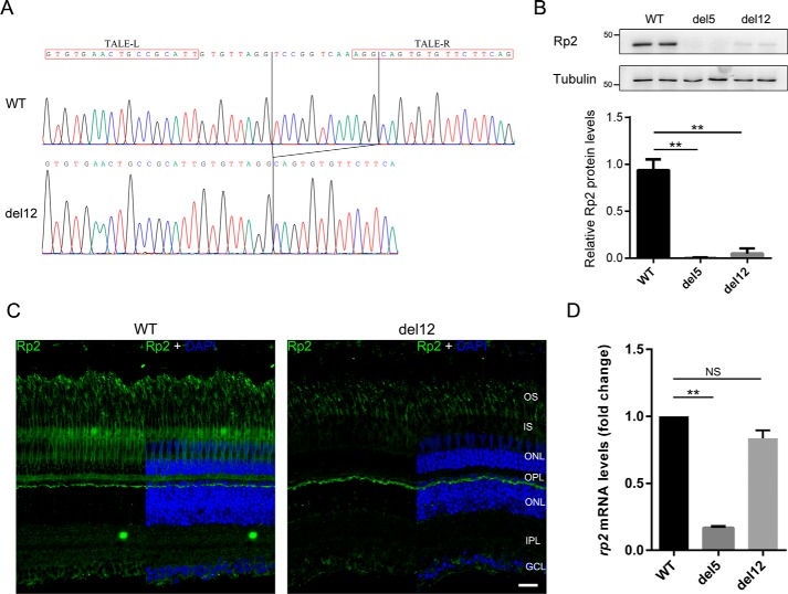
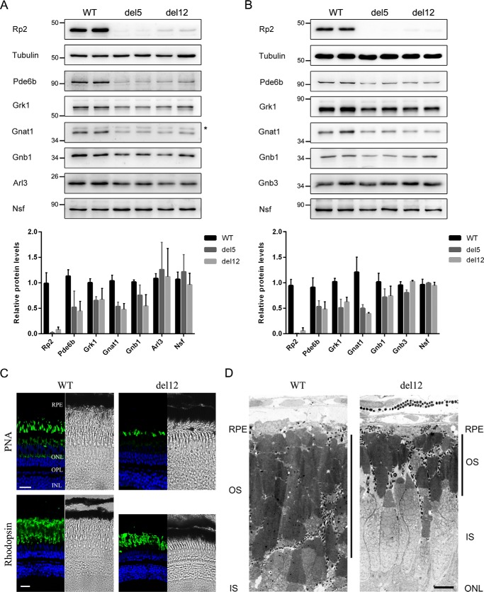
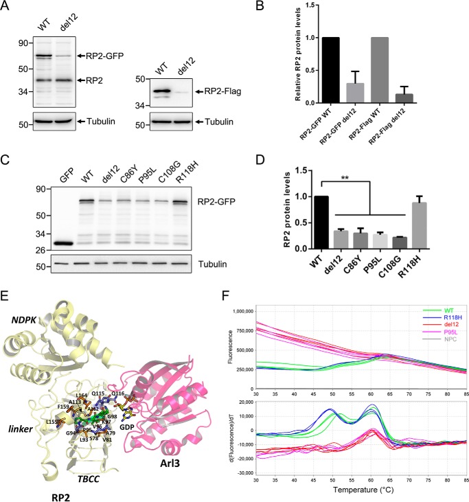
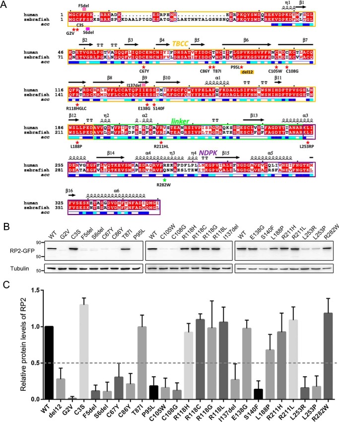
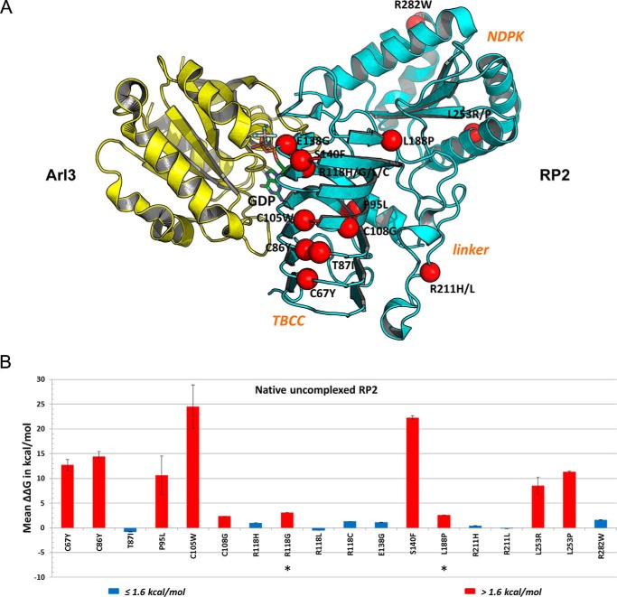
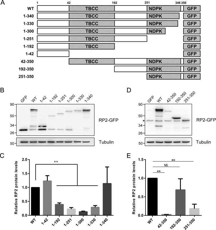
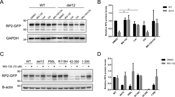
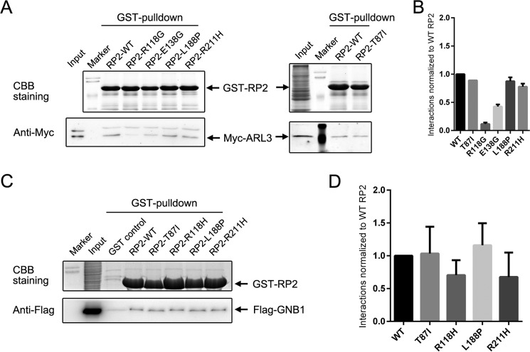
Mutations in retinitis pigmentosa 2 (RP2) account for 10-20% of X-linked retinitis pigmentosa (RP) cases. The encoded RP2 protein is implicated in ciliary trafficking of myristoylated and prenylated proteins in photoreceptor cells. To date >70 mutations in RP2 have been identified. How these mutations disrupt the function of RP2 is not fully understood. Here we report a novel in-frame 12-bp deletion (c.357_368del, p.Pro120_Gly123del) in zebrafish rp2 The mutant zebrafish shows reduced rod phototransduction proteins and progressive retinal degeneration. Interestingly, the protein level of mutant Rp2 is almost undetectable, whereas its mRNA level is near normal, indicating a possible post-translational effect of the mutation. Consistent with this hypothesis, the equivalent 12-bp deletion in human RP2 markedly impairs RP2 protein stability and reduces its protein level. Furthermore, we found that a majority of the RP2 pathogenic mutations (including missense, single-residue deletion, and C-terminal truncation mutations) severely destabilize the RP2 protein. The destabilized RP2 mutant proteins are degraded via the proteasome pathway, resulting in dramatically decreased protein levels. The remaining non-destabilizing mutations T87I, R118H/R118G/R118L/R118C, E138G, and R211H/R211L are suggested to impair the interaction between RP2 and its protein partners (such as ARL3) or with as yet unknown partners. By utilizing a combination of in silico, in vitro, and in vivo approaches, our work comprehensively indicates that loss of RP2 protein structural stability is the predominating pathogenic consequence for most RP2 mutations. Our study also reveals a role of the C-terminal domain of RP2 in maintaining the overall protein stability.

Keywords: RP2; mutant; protein degradation; protein stability; retinal degeneration; structural model; transcription activator-like effector nuclease (TALEN); zebrafish.


Conflict of interest statement

The authors declare that they have no conflicts of interest with the contents of this article

Figures

Figure 1.
Figure 1.
Identification of the del12 mutant zebrafish with dramatically decreased Rp2 protein levels. A, the c.357_368del mutation (del12) in zebrafish rp2 was confirmed by Sanger sequencing at the genomic level. The chromatograms of WT and del12 zebrafish are shown. The TALE binding sequences are marked by red boxes. The deleted 12 bp is labeled by vertical lines. B, protein levels of Rp2 were detected in WT, del5 (knock-out) and del12 mutant zebrafish at the age of 3 months by Western blot using the anti-zebrafish Rp2 antibody. Tubulin served as a loading control. Quantitative results of Rp2 protein levels in del5 and del12 homozygotes from three independent experiments (n = 6) are shown as the mean with S.D. in the lower panel. **, p < 0.01. C, immunostaining of Rp2 in dark-adapted retinas from WT and del12 mutant zebrafish at the age of 3 months. As compared with WT, the del12 mutants show much weaker fluorescent signals in the retina. OS, outer segment; IS, inner segment; ONL, outer nuclear layer; OPL, outer plexiform layer; INL, inner nuclear layer; IPL, inner plexiform layer, GCL, ganglion cell layer. Scale bar: 20 μm. D, the corresponding rp2 mRNA levels in 3-month-old WT and rp2 mutant zebrafish were detected by RT-qPCR. Actb1 was used as an endogenous control. The results are shown as the mean with S.D. (n = 3). **, p < 0.01. NS, not significant.
Figure 2.
Figure 2.
The del12 mutant zebrafish showed decreased rod phototransduction proteins and progressive retinal degeneration. A and B, protein levels of the rod phototransduction proteins (Pde6b, Grk1, Gnat1, and Gnb1) and cone phototransduction protein Gnb3 as well as the two RP2-interacting protein Arl3 and Nsf were detected by Western blot in 1-month-old (A) and 2-month-old (B) WT, del5, and del12 mutant zebrafish. Tubulin was used as a loading control. The asterisk in A indicates a nonspecific bands produced by the anti-GNAT1 antibody. The quantitative results are shown as the mean with S.D. (n = 6) in the lower panels, respectively. C, retinal cryosections from WT and del12 mutant zebrafish were stained with PNA (upper panel) and the anti-rhodopsin (4D2) antibody (lower panel) to label the outer segments of cones and rods at the age of 6 months. Reduction in the thickness of the rod outer segment layer is seen in del12 mutant zebrafish. Scale bars: 20 μm. D, retinal ultrastructure analysis of 7-month-old WT and del12 mutant zebrafish revealed significantly shortened outer segments of photoreceptors in del12 mutant zebrafish. RPE, retinal pigment epithelium; OS, outer segment; IS, inner segment; ONL, outer nuclear layer. Scale bars: 5 μm.
Figure 3.
Figure 3.
The del12 and nearby mutations in human RP2 decreased its protein level and stability. A, C-terminal GFP or FLAG-tagged WT and del12 mutant RP2 were transfected into ARPE-19 cells. The RP2 protein levels were detected by Western blot using the anti-human RP2 antibody or the anti-FLAG antibody. The endogenous and GFP or FLAG-tagged RP2 bands are indicated by arrows. Tubulin was used as a loading control. B, the quantitative results of A from five independent experiments are shown as the mean with S.D. (n = 5). The del12 mutation significantly reduced the protein levels of RP2. C, the nearby C86Y, P95L, and C108G mutations of RP2 show similar effects as the del12 mutation on the expression of RP2 in ARPE-19 cells. The R118H mutation was used as a control. GFP, the empty vector without RP2 sequence. D, the quantitative results of C from three independent experiments are shown as the mean with S.D. (n = 3). **, p < 0.01. E, intraprotein interactions of the four residues affected by the RP2 del12 mutation. Pro-95, Val-96, Lys-97, and Gly-98 are shown in stick representation colored green. Those residues that participate in H-bonds with the four residues are shown in blue, and hydrophobic interactions are in orange. F, the melting curve plots and derivative plots of WT, del12, P95L, and R118H forms of GST-RP2 fusion proteins in DSF analysis are shown. The high initial fluorescence signals of del12 and P95L groups (upper panel, red and purple lines) indicate that the proteins are partially or completely unfolded at the beginning. The two transitions (around 50 and 60 °C) of WT and R118H groups (lower panel, green and blue lines) represent the unfolding processes of RP2 and GST with increasing temperature, respectively. NPC, no protein control.
Figure 4.
Figure 4.
More than half of the RP2 missense mutations were destabilizing. A, the mutations investigated in this study are labeled in the alignment result of human and zebrafish RP2 protein sequences. Red star, missense mutations; pink filled rectangle, deletion mutations; yellow box, the del12 mutation; green star, polymorphism. Red background, strictly conserved positions; red letter with white background, conservatively substituted positions; black letter with white background, non-conserved positions. The secondary structures and solvent accessibility of each position are shown above and below the alignment. Blue, highly solvent-accessible; cyan, intermediate solvent-accessible; white, buried. B, 22 pathogenic forms of mutant RP2 proteins were expressed in ARPE-19 cells with a C-terminal GFP tag. Protein levels were detected by Western blot using the anti-GFP antibody. Tubulin was used as a loading control. The experiment shown was replicated at least three times. C, quantification of the data shown in B. The results are shown as mean with S.D. (n = 5). The protein levels of del12 and WT forms of RP2 are used as references to classify destabilizing and non-destabilizing mutations, respectively.
Figure 5.
Figure 5.
Structure-based modeling and analysis of RP2 mutations. A, missense mutations mapped on RP2-Arl3 crystal structure. A schematic representation of the RP2 (cyan)-Arl3 (yellow) complex is shown along with GDP (stick representation, colored by atom type). The location of known disease-causing missense mutations is shown on RP2. Only the positions of the α carbon atoms are shown as red spheres and labeled. B, analysis of RP2 missense mutations on structure-function by FoldX. The calculated ΔΔG (kcal/mol) is shown as the mean of three calculation runs with standard error. ΔΔG ≤ 1.6 kcal/mol, no effect on structural stability (blue); ΔΔG > 1.6 kcal/mol, severely reduced structural stability (red). Negative values indicate enhanced stabilities.
Figure 6.
Figure 6.
Expression levels of the C-terminal and N-terminal truncations of RP2. A, schematic diagram of the truncated RP2 proteins with a C-terminal GFP tag. The exact length of all studied truncations of RP2 is shown. B and D, the series of C-terminal truncated (B) and N-terminal truncated (D) RP2 were transfected into ARPE-19 cells, respectively. Their expression levels were determined by Western blot using either the anti-GFP antibody or the anti-RP2 antibody. Tubulin was used as a loading control. The experiment shown was replicated at least three times. C and E, the quantitative results of (B and D) are shown as the mean with S.D. (n = 3), correspondingly. **, p < 0.01; NS, non-significant.
Figure 7.
Figure 7.
The unstable RP2 proteins were degraded through proteasome pathway in ARPE-19 cells. A, ARPE-19 cells transfected with WT and del12 mutant RP2 were treated with MG-132 (10 μm), lactacystin (Lac, 20 μm), or chloroquine (Chl, 20 μm) for 18 h. The RP2 protein levels were detected by Western blot. GAPDH was used as a loading control. The experiment shown was repeated at least three times. B, quantitative analysis of the Western blot data shown in A revealed that the proteasome inhibitors MG-132 and lactacystin significantly increased the protein levels of del12 mutant RP2. The results are shown as the mean with S.D. n = 3. **, p < 0.01. C, the P95L, 42–350, and 1–300 forms of RP2 were transfected into ARPE-19 cells, which were further treated with MG-132 (10 μm) for 18 h. RP2 protein levels were detected by Western blot. GAPDH was used as a loading control. The experiment shown was repeated at least three times. D, quantitative results of C from three independent experiments are shown as the mean with S.D. (n = 3). MG-132 treatment markedly increased the protein levels of P95L and 1–300 forms of RP2 mutants.
Figure 8.
Figure 8.
GST pulldown assays of the non-destabilizing and partly destabilized RP2 mutations. A and C, pulldown results of WT and the indicated mutant forms of RP2 with Myc-tagged ARL3 (A) and FLAG-tagged GNB1 (C). Coomassie Brilliant Blue (CBB) staining shows the amounts of the immobilized GST-RP2 proteins. ARL3 and GNB1 are visualized by Western blot using corresponding antibodies. B and D, quantitative results of A and C are shown as the mean with S.D., respectively. The strength of the interaction is normalized to WT RP2 protein.

References

    1. Hartong D. T., Berson E. L., and Dryja T. P. (2006) Retinitis pigmentosa. Lancet 368, 1795–1809 - PubMed
    1. Prokisch H., Hartig M., Hellinger R., Meitinger T., and Rosenberg T. (2007) A population-based epidemiological and genetic study of X-linked retinitis pigmentosa. Invest. Ophthalmol. Vis. Sci. 48, 4012–4018 - PubMed
    1. Pelletier V., Jambou M., Delphin N., Zinovieva E., Stum M., Gigarel N., Dollfus H., Hamel C., Toutain A., Dufier J. L., Roche O., Munnich A., Bonnefont J. P., Kaplan J., and Rozet J. M. (2007) Comprehensive survey of mutations in RP2 and RPGR in patients affected with distinct retinal dystrophies: genotype-phenotype correlations and impact on genetic counseling. Hum. Mutat. 28, 81–91 - PubMed
    1. Li L., Khan N., Hurd T., Ghosh A. K., Cheng C., Molday R., Heckenlively J. R., Swaroop A., and Khanna H. (2013) Ablation of the X-linked retinitis pigmentosa 2 (Rp2) gene in mice results in opsin mislocalization and photoreceptor degeneration. Invest. Ophthalmol. Vis. Sci. 54, 4503–4511 - PMC - PubMed
    1. Zhang H., Hanke-Gogokhia C., Jiang L., Li X., Wang P., Gerstner C. D., Frederick J. M., Yang Z., and Baehr W. (2015) Mistrafficking of prenylated proteins causes retinitis pigmentosa 2. FASEB J. 29, 932–942 - PMC - PubMed

MeSH terms

LinkOut - more resources