. 2017 Apr 28;120(9):1403-1413.
doi: 10.1161/CIRCRESAHA.116.308741. Epub 2017 Feb 16.

Demethylation of H3K27 Is Essential for the Induction of Direct Cardiac Reprogramming by miR Combo

Affiliations

Demethylation of H3K27 Is Essential for the Induction of Direct Cardiac Reprogramming by miR Combo

Sophie Dal-Pra et al. Circ Res. .

Abstract

Rationale: Direct reprogramming of cardiac fibroblasts to cardiomyocytes has recently emerged as a novel and promising approach to regenerate the injured myocardium. We have previously demonstrated the feasibility of this approach in vitro and in vivo using a combination of 4 microRNAs (miR-1, miR-133, miR-208, and miR-499) that we named miR combo. However, the mechanism of miR combo mediated direct cardiac reprogramming is currently unknown.

Objective: Here, we investigated the possibility that miR combo initiated direct cardiac reprogramming through an epigenetic mechanism.

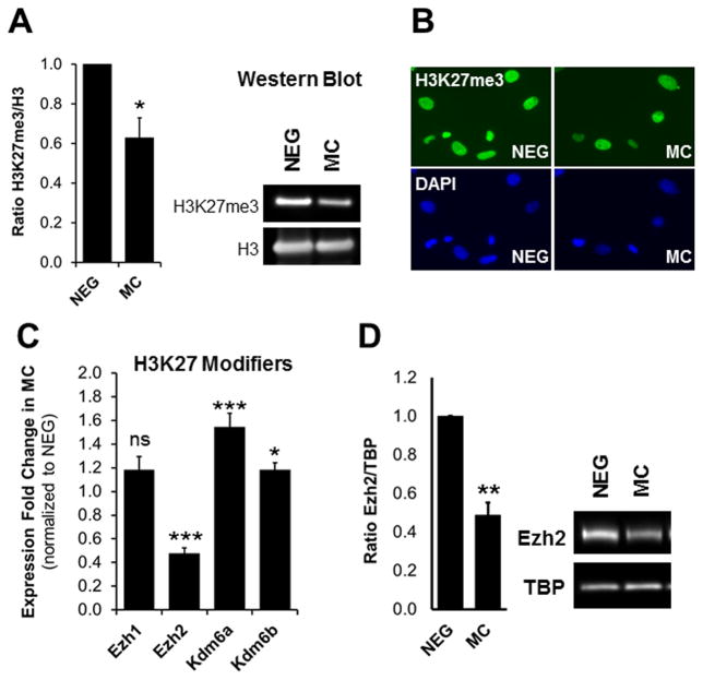
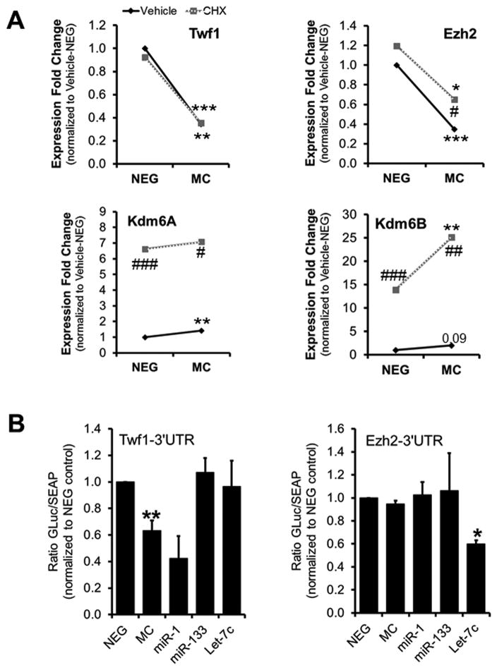
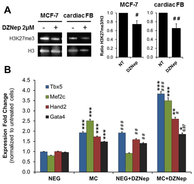
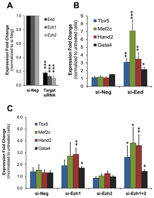
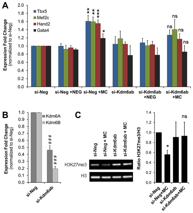
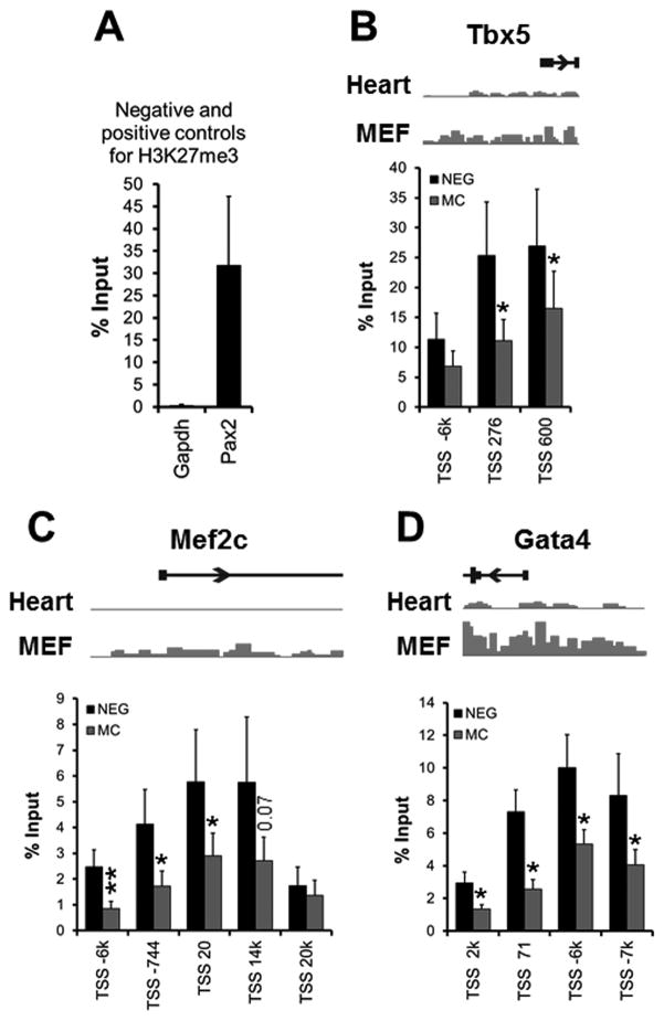
Methods and results: Using a quantitative polymerase chain reaction array, we found that histone methyltransferases and demethylases that regulate the trimethylation of H3K27 (H3K27me3), an epigenetic modification that marks transcriptional repression, were changed in miR combo-treated fibroblasts. Accordingly, global H3K27me3 levels were downregulated by miR combo treatment. In particular, the promoter region of cardiac transcription factors showed decreased H3K27me3 as revealed by chromatin immunoprecipitation coupled with quantitative polymerase chain reaction. Inhibition of H3K27 methyltransferases or of the PRC2 (Polycomb Repressive Complex 2) by pharmaceutical inhibition or siRNA reduced the levels of H3K27me3 and induced cardiogenic markers at the RNA and protein level, similarly to miR combo treatment. In contrast, knockdown of the H3K27 demethylases Kdm6A and Kdm6B restored the levels of H3K27me3 and blocked the induction of cardiac gene expression in miR combo-treated fibroblasts.

Conclusions: In summary, we demonstrated that removal of the repressive mark H3K27me3 is essential for the induction of cardiac reprogramming by miR combo. Our data not only highlight the importance of regulating the epigenetic landscape during cell fate conversion but also provide a framework to improve this technique.

Keywords: cardiac myocytes; cellular reprogramming; chromatin; epigenomics; microRNAs; regeneration.


Figures

Figure 1
Figure 1. MiR combo treatment affects H3K27 methylation and the expression of H3K27 modifiers
(A) Analysis of H3K27me3 levels in negmiR (NEG) and miR combo (MC) treated neonatal cardiac fibroblasts by immunoblot. The graph presents the quantification of H3K27me3 levels normalized to histone H3 levels (N=3). (B) Analysis of H3K27me3 levels in NEG- and MC- treated neonatal cardiac fibroblasts by immunocytochemistry (N=3). (C) Analysis of H3K27 methyltransferase (Ezh1 and Ezh2) and demethylase (Kdm6A and Kdm6B) mRNA levels in MC-treated neonatal fibroblasts by qPCR, 3 days after transfection. Expression data was normalized to NEG-treated cells (N=7). (D) Analysis of Ezh2 protein levels by immunoblot in neonatal cardiac fibroblasts transfected with NEG or MC. TBP was used as loading control and Ezh2/TBP ratios were normalized to NEG controls (N=3). Asterisks indicate statistical significance between NEG and MC-treated cells determined by standardized T-test (*P ≤0.01, **P ≤0.005 or ***P≤0.0005).
Figure 2
Figure 2. Regulation of the expression of H3K27 modifiers by miR combo
(A) Analysis of the mRNA levels of Twf1, Ezh2, Kdm6A and Kdm6B by qPCR in NEG or MC transfected cells treated for 12 hours with vehicle (DMSO) or 50 μg/ml of cycloheximide (CHX) to block protein synthesis (N=3–4). (B) Luciferase reporter assays were performed in HEK293 cells by co-transfecting Twf1-3′-UTR vector or Ezh2-3′-UTR vector with negmiR (NEG), miR combo (MC), miR-1, miR-133 or Let7c. The Twf1-3′UTR reporter was used as a positive control for miR combo (Twf1 is a direct target of miR-1). Let7c was used as a positive control for Ezh2-3′UTR (which contains the predicted Let7c binding sequence). Comparisons were made between NEG and MC (*P ≤0.05, **P ≤0.01 or ***P≤0.005) or between CHX and vehicle-treated cells (# P≤0.05, ## P ≤0.01 or ### P ≤0.005) using paired T-test.
Figure 3
Figure 3. Inhibition of H3K27 methylation induces cardiac transcription factor expression
(A) Analysis of H3K27me3 levels in MCF7 and neonatal cardiac fibroblasts (FB) cultured for 3 days in growth media containing 2 μmol/L of DZNep or the vehicle (N=3). Immunoblots and quantification are shown as indicated. The H3K27me3/H3 ratio in DZNep treated cells was normalized to the vehicle. (B) Neonatal cardiac fibroblasts were transfected with negmiR (NEG) or miR combo (MC) and cultured in the presence of 2 μmol/L of DZNep or vehicle for 3 days. Tbx5, Mef2c, Hand2 and Gata4 mRNA levels were determined by qPCR (N≥8). Comparisons were made between NEG and MC (*P ≤0.05, **P ≤0.01 or ***P≤0.0005) or between DZNep and vehicle-treated cells (# P≤0.01, ## P ≤0.005 or ### P ≤0.0005) using a standardized T test
Figure 4
Figure 4. Knockdown of PRC2 complex members induces cardiac transcription factor expression
(A) Expression levels of Eed, Ezh1 and Ezh2 in neonatal cardiac fibroblasts transfected with negative siRNA (si-Neg) or siRNA against Eed (si-Eed), Ezh1 (si-Ezh1) or Ezh2 (si-Ezh2). mRNAs were isolated 3 days after transfection and analyzed by qPCR. Expressions were normalized to si-Neg (N=5). (B and C) Cardiac transcription factor expression was analyzed by qPCR in neonatal cardiac fibroblasts transfected with si-Neg, si-Eed (B), si-Ezh1, si-Ezh2 or si-Ezh1+2 (C), 3 days after transfection. The data was normalized to the si-Neg control (N=5–6). For all panels, comparisons were made between si-Neg and target siRNA (*P ≤0.05, **P ≤0.01 or ***P ≤0.005).
Figure 5
Figure 5. Inhibition of H3K27me3 demethylases blocks the induction of cardiac reprogramming by miR combo
(A) Analysis of the expression of cardiac transcription factors in neonatal cardiac fibroblasts transfected with negmiR (NEG) or miR combo (MC), in the presence of negative siRNA (si-Neg) or siRNA targeting Kdm6A and Kdm6B (si-Kdm6A+B). mRNA were isolated 3 days after transfection and analyzed by qPCR. Expression values were normalized to the si-Neg control. (N=5). (B) mRNA levels of Kdm6A and Kdm6B in neonatal cardiac fibroblasts transfected with si-Kdm6A+B compared to control fibroblasts transfected with si-Neg. (N=5). (C) Immunoblot analysis of H3K27me3 levels in neonatal cardiac fibroblasts treated with NEG or MC in combination with si-Neg or si-Kdm6A+B, 3 days after transfection (N=3). For all panels, comparisons were made between NEG and MC ((*p ≤0.05, **p ≤0.005 or ***p ≤0.0005) or between si-Neg and si-Kdm6A+B (### P≤ 0.0005) respectively.
Figure 6
Figure 6. Reduction of H3K27me3 at loci of genes encoding cardiac transcription factors
(A) Analysis of the negative (gapdh) and positive (pax2) controls for H3K27me3, by ChIP-qPCR for H3K27me3 in neonatal cardiac fibroblasts. (B-D) Analysis of the promoter region of Tbx5 (B), Mef2c (C) and Gata4 (D) by ChIP-qPCR for H3K27me3 (N=6). Chromatin samples were isolated from neonatal cardiac fibroblasts transfected with negmiR (NEG) or miR combo (MC) and processed 3 days after transfection. Results were expressed as a percentage of the input control. For each gene, we used a set of 3 to 5 validated primer pairs which were named according to their location relative to the transcription start (TSS). For each gene, a schematic representation of their H3K27me3 profile in heart and in fibroblasts (MEF) is shown. Comparisons were made between NEG and MC (*p ≤0.05) using 2-tails paired T-test.

Comment in

References

    1. Lin Z, Pu WT. Strategies for cardiac regeneration and repair. Sci Transl Med. 2014;6:239rv1. - PMC - PubMed
    1. Xin M, Olson EN, Bassel-Duby R. Mending broken hearts: cardiac development as a basis for adult heart regeneration and repair. Nat Rev Mol Cell Biol. 2013;14:529–41. - PMC - PubMed
    1. Dal-Pra S, Mirotsou M. Reprogramming approaches in cardiovascular regeneration. Curr Treat Options Cardiovasc Med. 2014;16:327. - PubMed
    1. Hodgkinson CP, Kang MH, Dal-Pra S, Mirotsou M, Dzau VJ. MicroRNAs and Cardiac Regeneration. Circ Res. 2015;116:1700–11. - PMC - PubMed
    1. Sadahiro T, Yamanaka S, Ieda M. Direct cardiac reprogramming: progress and challenges in basic biology and clinical applications. Circ Res. 2015;116:1378–91. - PubMed

MeSH terms