. 2017 Mar 28;8(13):20813-20824.
doi: 10.18632/oncotarget.15343.

Exclusive destruction of mitotic spindles in human cancer cells

Affiliations

Exclusive destruction of mitotic spindles in human cancer cells

Leonid Visochek et al. Oncotarget. .

Erratum in

Abstract

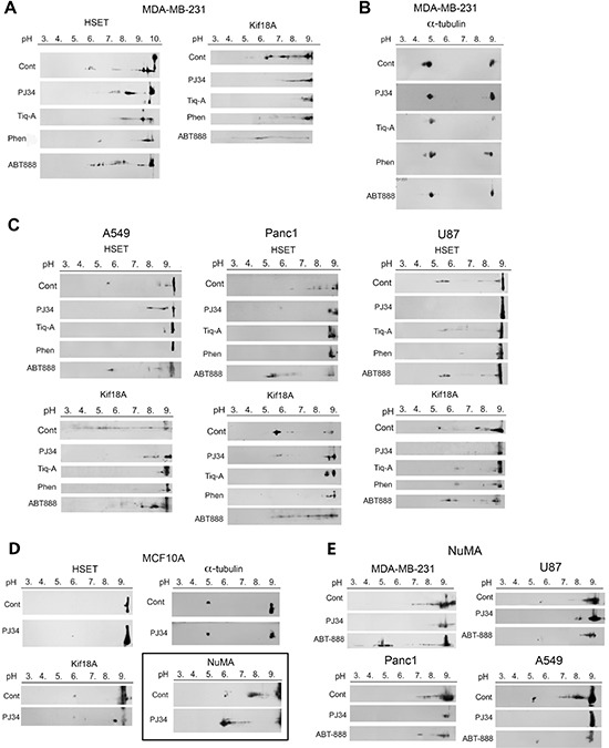
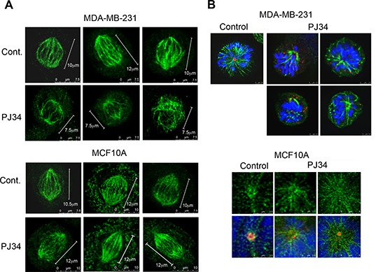
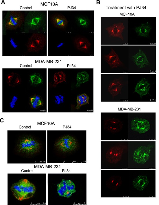
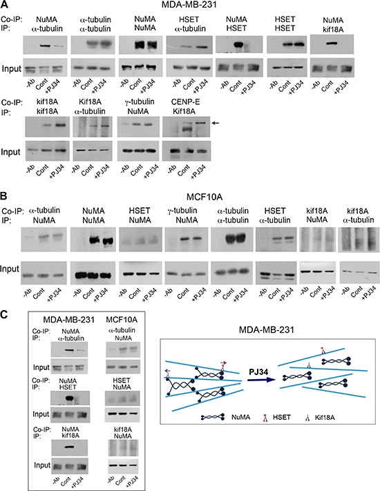
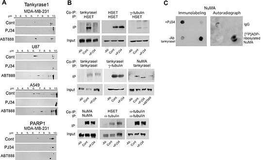
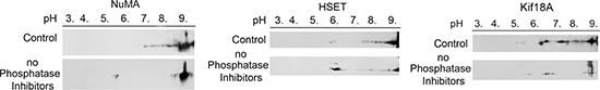
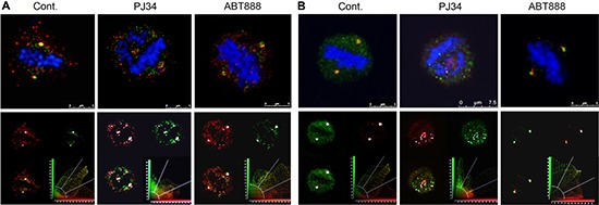
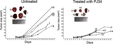
We identified target proteins modified by phenanthrenes that cause exclusive eradication of human cancer cells. The cytotoxic activity of the phenanthrenes in a variety of human cancer cells is attributed by these findings to post translational modifications of NuMA and kinesins HSET/kifC1 and kif18A. Their activity prevented the binding of NuMA to α-tubulin and kinesins in human cancer cells, and caused aberrant spindles. The most efficient cytotoxic activity of the phenanthridine PJ34, caused significantly smaller aberrant spindles with disrupted spindle poles and scattered extra-centrosomes and chromosomes. Concomitantly, PJ34 induced tumor growth arrest of human malignant tumors developed in athymic nude mice, indicating the relevance of its activity for cancer therapy.

Keywords: NuMA; human cancer cells; kinesins; mitotic spindles; phenanthrenes.


Conflict of interest statement

CONFLICTS OF INTEREST

The authors declare no competing financial interests. M.C-A. is the inventor in patents US 8,729,080 B2 and EP 2 207 548B1.

Figures

Figure 1
Figure 1. Post-translational modifications of kinesins and NuMA identified by changes in their isoelectric point in human cells treated with phenanthrenes
(A) The isoelectric point of kinesins HSET/kifC1 and kif18A in human triple negative breast cancer cells MDA-MB-231 was shifted towards higher pH values (less negatively charged protein) after 27 hours incubation with the phenanthrene derivatives PJ34 (20 μM), Tiq-A (50 μM) or Phen (50 μM). No similar effect was induced by the non-phenanthrene PARP1 inhibitor ABT888 (20 μM). (B) The isoelectric point of α-tubulin was not affected by the phenanthrenes or by ABT888 in MDA-MB-231 cells. (C) The isoelectric point of kinesins HSET/kifC1 and kif18A was shifted towards higher pH values (less negatively charged protein) in human lung (A549), pancreas (PANC1) and glioblastoma (U87) cancer cells incubated for 27 h with PJ34 (20 μM), Tiq-A (50 μM) or Phen (50 μM). No similar effect on their isoelectric point was induced by the non-phenanthrene PARP1 inhibitor ABT888 (20 μM). (D) Treatment with the phenanthridine PJ34 (20 μM, 27 h) did not affect the isoelecrtic point of α-tubulin and kinesins HSET/kifC1 and Kif18A in human normal breast epithelial cells MCF10A. The isoelectric point of NuMA was shifted towards lower pH (more negatively charged protein) in these cells. (E) Treatment with PJ34 (20 μM, 27 h) shifted the isoelectric point of NuMA towards higher pH values (less negatively charged protein) in human malignant cells, including: breast (MDA-MB-231), lung (A549), pancreas (PANC1) and glioblastoma (U87) cells. Representative results of 4 different experiments in each cell type are displayed in A–E.
Figure 2
Figure 2. Aberrant spindles and impaired spindle poles in human cancer cells treated with the phenanthridine PJ34
(A) Small aberrant spindles were identified by confocal microscopy in randomly scanned human breast cancer MDA-MB-231 cells incubated with PJ34 (20 μM, 27 h). Spindles were immunolabeled for HSET/kifC1 (green) bound to microtubules. Similarly immunolabeled spindles are not impaired in PJ34 treated normal human breast epithelial cells MCF10A. Bars indicate the spindle length measured by the scale bar (n = 20; 3 different experiments), 95% of the spindles in MDA-MB-231 cells were shorter, 60 ± 5% in size. (B) Upper panel: Impaired spindle poles were identified in randomly scanned fixed MDA-MB-231 cells treated with PJ34 (20 μM, 27 h). Microtubules immunolabeled by α- tubulin (green), centrosomes immunolabeled by γ-tubulin (red) and chromosomes stained by DAPI (blue) are displayed. Lower panel: Spindle poles were not impaired in randomly scanned normal epithelial MCF10A cells treated with PJ34 (20 μM, 27 h). Microtubules identified by immunolabeled HSET14/kifC1 (green), centrosomes immunolabeled for γ-tubulin (red) and chromosomes stained by DAPI (blue) are displayed. The sampled spindles represent 95% of randomly scanned spindles (n = 20) of cancer MDA-MB-231 cells and all the randomly scanned spindles (n = 20) of normal epithelial MCF10A cells in 3 different experiments.
Figure 3
Figure 3. Impaired NuMA alignment in impaired spindles of human cancer cells treated with the phenanthridine PJ34
Scanned spindles in fixed human breast cancer MDA-MB-231 cells (upper frames in A and B) and normal breast epithelial MCF10A cells (lower frames in A and B), without or following treatment with PJ34 (20 μM, 27 h). Aberrant disorganized spindles, impaired NuMA alignment, and impaired chromosome alignments in the spindle mid-zone were observed only in cancer cells treated with PJ-34. Normal and malignant cells were immunolabeled for NuMA (red) and α-tubulin (green). Chromosomes are stained with DAPI (blue). (C) Scattered misaligned chromosomes and NuMA in disorganized spindles following PJ34 treatment (20 μM, 27 h) of MDA-MB-231 cancer cells. Proper alignment of chromosomes in the mid-zone of bifocal spindles and NuMA labeling in bifocal poles of PJ34 treated (20 μM, 27 h) MCF10A human epithelial cells. Both cell types were immunolabeled for NuMA (red) and kinesin HSET/kifC1 (green). Chromosomes are stained with DAPI (blue). Disorganized spindles and misaligned chromosomes were observed in 95% of spindles in PJ34 treated MDA-MB-231 cells (n = 20). Normal spindles, and chromosomes aligned in the spindle mid-zone were observed in all scanned PJ34 treated MCF10A cells (n = 20).
Figure 4
Figure 4. An impaired binding of NuMA to α-tubulin and to kinesins HSET/kifC1 and kif18A in human cancer cells treated with PJ34
NuMA was not co-immunoprecipitated with α-tubulin or kinesins HSET/kifC1 and kif18A in breast cancer cells MDA-MB-231 treated with PJ34 (20 μM, 27 h) (A and Supplementary Figure 2). No impaired binding of NuMA to α-tubulin and kinesins was detected in similarly treated human breast epithelial cells MCF10A (B). These results are schematically displayed in (C). Average 11.3 ± 1.1 times reduction in co-immunoprecipitated NuMA with α-tubulin was measured by scanning in PJ34 treated versus untreated MDA-MB-231 cancer cells. Average 1.2 ± 0.2 times reduction in the co-immunoprecipitated NuMA with α-tubulin was measured by scanning in PJ34 treated versus untreated normal epithelial cells MCF10A. Representative results of 4 different experiments are displayed.
Figure 5
Figure 5. PJ34 inhibits NuMA and tankyrase1 polyADP-ribosylation in cancer cells
(A) Inhibition of tankyrase1 modification measured by its shifted pI. Treatment with PJ34 (20 μM, 27 h) attenuated the shift in the isoelectric point of negatively charged tankyrase1 in lung cancer (A549), triple negative breast cancer (MDA-MB-231) and glioblastoma (U87) cancer cells. No similar shift of the isoelectric point of tankyrase1 was induced by the non-phenanthrene PARP1 inhibitor ABT888 (20 μM, 27 h). PolyADP-ribosylated PARP1 was similarly suppressed by both PARP1 inhibitors, PJ34 (20 μM) and ABT888 (20 μM). Representative results of 3 experiments in each cell type are displayed. (B) The binding of tankyrase1 to γ-tubulin or NuMA was measured by co-immunoprecipitation. Their binding in MDA-MB-231 cells was not affected by treatment with PJ34 (20 μM, 27 h). The binding of tankyrase1 to kinesin HSET/kifC1 was impaired. Representative results of 3 experiments are displayed. (C) [32P]polyADP-ribosylation of NuMA bound to tankyrase1 was inhibited after treatment of human cancer cells MDA-MB-231 with PJ34 (20 μM, 30 min). Binding of NuMA to tankyrase1 was measured by dot blot analysis (Methods). [32P]polyADP-ribosylated NuMA bound to tankyrase1 was trapped by tankyrase1 antibody on nitrocellulose membrane, auto-radiographed and immunolabeled. Results were repeated in 3 different experiments.
Figure 6
Figure 6. Phosphatase activity changes the isoelectric points of NuMA and kinesins HSET/kifC1 and kif18A in human breast cancer cells MDA-MB-231
In the absence of phosphatase inhibition enhancing kinase activity, some of the isoelectric points of NuMA and kinesins HSET/kifC1 and kif18A were shifted towards higher pH values (less negatively charged protein). Similar results were obtained in 3 different experiments.
Figure 7
Figure 7. Co-localization of tankyrase1 with γ-tubulin and pericentrin in spindles of multi-centrosomal MDA-MB-231 cancer cells
Bi-polar co-localization of tankyrase1 and γ-tubulin (A) or tankyrase1 and pericentrin (B) in spindles of untreated MDA-MB-231 cancer cells were identified by confocal microscopy scanning. Fixed cultured MDA-MB-231 cells were immunolabeled for tankyrase1 (green), γ-tubulin (red) and pericentrin (red). Chromosomes are stained by DAPI (blue). After incubation with PJ34 (20 μM, 27 h), the bipolar co-localization of tankyrase1 and γ-tubulin (A) or tankyrase1 and pericentrin (B) turned into multiple scattered foci of the co-localized proteins. Chromosomes were not aligned in the spindle mid-zone in all the PJ34 treated cells. Treatment with ABT888 (n = 20; 20 μM, 27 h) did not impair the bipolar co-localization of tankyrase1 and γ-tubulin (A), or tankyrase and pericentrin (B), nor impaired the chromosomes alignment in the spindle mid-zone in MDA-MB-231 cells. Co-localization of tankyrase1 and pericentrin, or tankyrase1 and γ-tubulin was detected in 95% of the randomly scanned spindles of both treated and untreated cells (n = 20).
Figure 8
Figure 8. Human tumors growth arrested in nude athymic mice treated with PJ34
The growth of human triple negative breast MDA-MB-231 xenotransplants was significantly arrested in the indicated nude athymic mice injected i.p. (intraperitoneal) with PJ34 (60 mg/kg, daily for 14 days). The significance of the change in tumor growth rate was calculated by using one-tailed t-test for estimating the statistical significance of the difference between the group means, without assuming equal variance. The result was statistically significant: p = 0.036 (t = 2.14, df = 6.33). The daily measured average tumor size in each group is presented in Supplementary Figure 3.

References

    1. Bryant HE, Schultz N, Thomas HD, Parker KM, Flower D, Lopez E, Kyle S, Meuth M, Curtin NJ, Helleday T. Specific killing of BRCA2-deficient tumours with inhibitors of poly(ADP-ribose) polymerase. Nature. 2005;434:913–917. - PubMed
    1. Farmer H, McCabe N, Lord CJ, Tutt AN, Johnson DA, Richardson TB, Santarosa M, Dillon KJ, Hickson I, Knights C, Martin NM, Jackson SP, Smith GC, et al. Targeting the DNA repair defect in BRCA mutant cells as a therapeutic strategy. Nature. 2005;434:917–921. - PubMed
    1. Mangerich A, Bürkle A. How to kill tumor cells with inhibitors of poly(ADP-ribosyl)- ation. Int J Cancer. 2011;128:251–265. - PubMed
    1. Jagtap P, Szabo C. Poly(ADP-ribose)polymerase and the therapeutic effects of its inhibitors. Nat Rev Drug Discovery. 2005;4:421–440. - PubMed
    1. Passeri D, Camaioni E, Liscio P, Sabbatini P, Ferri M, Carotti A, Giacchè N, Pellicciari R, Gioiello A, Macchiarulo A. Concepts and Molecular Aspects in the Polypharmacology of PARP-1 Inhibitors. ChemMedChem. 2016;11:1219–1226. - PubMed

MeSH terms