. 2017 Feb 16;9(2):556-572.
doi: 10.18632/aging.101178.

Characterization of the influence of age on GABAA and glutamatergic mediated functions in the dorsolateral prefrontal cortex using paired-pulse TMS-EEG

Affiliations

Characterization of the influence of age on GABAA and glutamatergic mediated functions in the dorsolateral prefrontal cortex using paired-pulse TMS-EEG

Yoshihiro Noda et al. Aging (Albany NY). .

Abstract

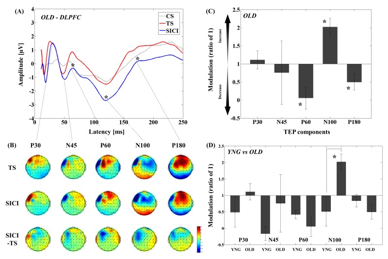
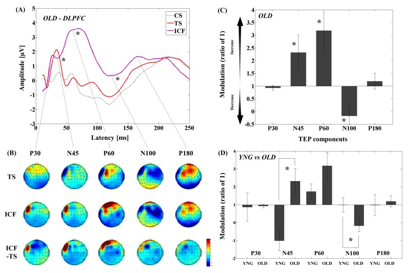
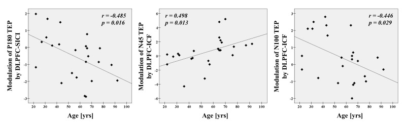
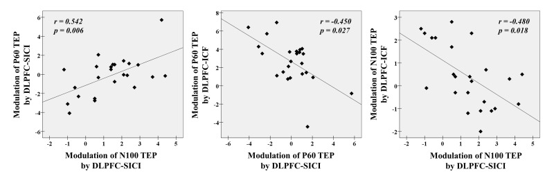
Gamma-aminobutyric acid (GABA)ergic and glutamatergic neurotransmissions in the prefrontal cortex decreases with age. Further, cognitive function mediated through the dorsolateral prefrontal cortex (DLPFC) also declines with age. Although neuroimaging studies have demonstrated decreased levels of these substances, direct neurophysiological data investigating the effect of aging in the DLPFC in human subjects is lacking. The advent of transcranial magnetic stimulation (TMS) combined with electroencephalography (EEG) has allowed for the assessment of functional neurotransmission in vivo. In the present study, we examined short interval intracortical inhibition (SICI) and intracortical facilitation (ICF) in a group of older adults (> 60 yrs) to evaluate the strength of GABAA and glutamate-mediated neurotransmission in the DLPFC, compared to younger adults (18-59 yrs). Older adults showed an increase of amplitude of N100 by the SICI paradigm, while N45 amplitude was increased and N100 amplitude was decreased by ICF. Moreover, these modulations significantly correlated with age. Our findings provide evidence for age-related alterations of excitatory and inhibitory functions in the prefrontal cortex in healthy adults. Future studies may aim to explore these neurophysiological relationships in the DLPFC in pathological forms of aging that affect cortical functioning such as mild cognitive impairment and Alzheimer's disease.

Keywords: TMS–EEG; aging effect; dorsolateral prefrontal cortex (DLPFC); intracortical facilitation (ICF); short interval intracortical inhibition (SICI).


Conflict of interest statement

CONFLICTS OF INTEREST

None of the authors declare any conflict of interest.

Figures

Figure 1
Figure 1. Modulation of TEPs by the DLPFC-SICI paradigm in older adults
(A) The graph depicts TEP traces averaged across the older adults for subthreshold TMS (black dot line: CS), unconditioned TMS (red line: TS) and conditioned TMS (blue line: SICI; ISI = 2ms) at the left frontal ROI. (B) The illustration shows the EEG topographical plots for conditions of TS alone, SICI, and the difference between TS and SICI obtained from the DLPFC-SICI experiment. Each vertical column depicts the TEP topoplots for P30, N45, P60, N100, and P180 component from left to right. (C) The bar graph shows modulatory effects of the DLPFC-SICI on TEPs in the older adults. The ANOVA and post-hoc analyses revealed that there are significant modulations (p < 0.05) in P60, N100, and P180 TEPs with the DLPFC-SICI paradigm. (D) The bar graph showing cross-sectional comparisons between younger and older adults in the DLPFC–SICI paradigm. The older adults show a significant facilitation of amplitude of N100 TEP than the younger adults (p < 0.05).
Figure 2
Figure 2. Modulation of TEPs by the DLPFC-ICF paradigm in older adults
(A) The graph depicts TEP traces averaged across the older adults for subthreshold TMS (black dot line: CS), unconditioned TMS (red line: TS) and conditioned TMS (purple line: ICF; ISI = 10ms) at the left frontal ROI. (B) The illustration shows the EEG topographical plots for conditions of TS alone, ICF, and the difference between TS and ICF obtained from the DLPFC-ICF experiment. Each vertical column depicts the TEP topoplots for P30, N45, P60, N100, and P180 component from left to right. (C) The bar graph shows modulatory effects of the DLPFC-ICF on TEPs in the older adults. The ANOVA and post-hoc analyses revealed that there are significant modulations (p < 0.05) in N45, P60, and N100 TEPs with the DLPFC-ICF paradigm. (D) The bar graph showing cross-sectional comparisons between younger and older adults in the DLPFC–ICF paradigm. The older adults demonstrate a significant facilitation of amplitude on N45 TEP and a significant attenuation of amplitude on N100 TEP compared to the younger adults (p < 0.05).
Figure 3
Figure 3. Age-related modulations on TEPs in all participants
There are significant correlations between age and modulation of P180 TEP by DLPFC-SICI (r = -0.485, p = 0.016, N = 24), as well as between age and modulations of N45 (r = 0.498, p = 0.013, N = 24) and N100 TEPs by DLPFC-ICF (r = -0.446, p = 0.029, N = 24) at the left frontal ROI.
Figure 4
Figure 4. Neurophysiological relationship with SICI and ICF paradigms from the DLPFC
There are significant correlations between modulations of P60 TEP and N100 TEP with DLPFC-SICI (r = 0.542, p = 0.006, N = 24), and further between a modulation of P60 TEP by DLPFC-SICI and a modulation of P60 TEP by DLPFC-ICF (r = -0.450, p = 0.027, N = 24) and between a modulation of N100 TEP by DLPFC-SICI and a modulation of N100 TEP by DLPFC-ICF (r = -0.480, p = 0.018, N = 24) at the left frontal ROI.

References

    1. Kaiser LG, Schuff N, Cashdollar N, Weiner MW. Age-related glutamate and glutamine concentration changes in normal human brain: 1H MR spectroscopy study at 4 T. Neurobiol Aging. 2005;26:665–72. doi: 10.1016/j.neurobiolaging.2004.07.001. - DOI - PMC - PubMed
    1. Grachev ID, Apkarian AV. Aging alters regional multichemical profile of the human brain: an in vivo 1H-MRS study of young versus middle-aged subjects. J Neurochem. 2001;76:582–93. doi: 10.1046/j.1471-4159.2001.00026.x. - DOI - PubMed
    1. Peinemann A, Lehner C, Conrad B, Siebner HR. Age-related decrease in paired-pulse intracortical inhibition in the human primary motor cortex. Neurosci Lett. 2001;313:33–36. doi: 10.1016/S0304-3940(01)02239-X. - DOI - PubMed
    1. Oliviero A, Profice P, Tonali PA, Pilato F, Saturno E, Dileone M, Ranieri F, Di Lazzaro V. Effects of aging on motor cortex excitability. Neurosci Res. 2006;55:74–77. doi: 10.1016/j.neures.2006.02.002. - DOI - PubMed
    1. Kossev AR, Schrader C, Däuper J, Dengler R, Rollnik JD. Increased intracortical inhibition in middle-aged humans; a study using paired-pulse transcranial magnetic stimulation. Neurosci Lett. 2002;333:83–86. doi: 10.1016/S0304-3940(02)00986-2. - DOI - PubMed

Publication types