Review
. 2017 Feb 2:10:22.
doi: 10.3389/fnmol.2017.00022. eCollection 2017.

APP as a Protective Factor in Acute Neuronal Insults

Affiliations
Review

APP as a Protective Factor in Acute Neuronal Insults

Dimitri Hefter et al. Front Mol Neurosci. .

Abstract

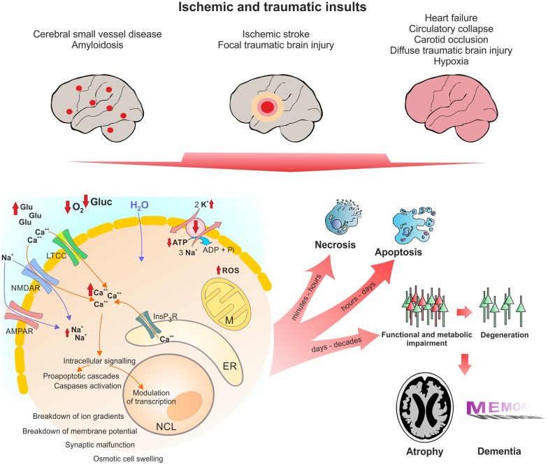
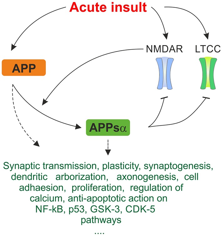
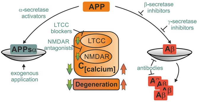
Despite its key role in the molecular pathology of Alzheimer's disease (AD), the physiological function of amyloid precursor protein (APP) is unknown. Increasing evidence, however, points towards a neuroprotective role of this membrane protein in situations of metabolic stress. A key observation is the up-regulation of APP following acute (stroke, cardiac arrest) or chronic (cerebrovascular disease) hypoxic-ischemic conditions. While this mechanism may increase the risk or severity of AD, APP by itself or its soluble extracellular fragment APPsα can promote neuronal survival. Indeed, different animal models of acute hypoxia-ischemia, traumatic brain injury (TBI) and excitotoxicity have revealed protective effects of APP or APPsα. The underlying mechanisms involve APP-mediated regulation of calcium homeostasis via NMDA receptors (NMDAR), voltage-gated calcium channels (VGCC) or internal calcium stores. In addition, APP affects the expression of survival- or apoptosis-related genes as well as neurotrophic factors. In this review, we summarize the current understanding of the neuroprotective role of APP and APPsα and possible implications for future research and new therapeutic strategies.

Keywords: Alzheimer; amyloid precursor protein; calcium toxicity; cell death; ischemia; neuroprotection; stroke; traumatic brain injury.


Figures

Figure 1
Figure 1
Pathophysiological changes in neurons following acute ischemic and traumatic insults. Micro- and macroscopic focal strokes, global hypoxia-ischemia and traumatic brain injury (TBI) lead to abruption of extracellular glucose and oxygen supply and excessive glutamate release. One major shared pathomechanism is NMDAR-mediated excitotoxicity, or over-activation of NMDAR by glutamate, which facilitates sodium and calcium influx. Due to excessive ion influx, the cellular membrane potential is depolarized, which leads to activation of voltage gated calcium channels such as LTCC, initiating a vicious cycle of ion influx, calcium overload, depolarization and aberrant activity. Successively calcium from intracellular calcium stores, particularly mitochondria and the ER, is released, increasing calcium levels to up to 200-fold of ~100 nM during resting. Calcium activates secondary messengers that are able to translocate to the nucleus and modulate gene transcription. Long-lasting or severely elevated calcium levels may lead to activation of caspases and apoptosis. Following the osmotic gradient, water enters the cell and leads to cell swelling and brain edema. Due to glucose and oxygen shortage and excessive formation of reactive oxygen species (ROS), mitochondrial function is compromised and ATP production halts. Malfunction of the energy demanding ion pumps, predominantly the sodium potassium pump, ultimately leads to breakdown of the membrane potential, a phenomenon known as anoxic or hypoxic spreading depolarization or spreading depression (due to depression of network activity in the field potential recording). Given the energy supply is timely restored, this stage can be reversed without long-lasting morphological damage. If the insult is protracted, neurons might undergo (dependent of insult’s severity) necrotic or apoptotic death or degenerate with a delay of days to decades due to synaptic or metabolic malfunction. Acute cell death and delayed degeneration contribute to brain atrophy and development of dementia. Glu, glutamate; Gluc, glucose; LTCC, L-Type calcium channel; NMDAR, NMDA receptor; AMPAR, AMPA receptor; M, mitochondrion; NCL, nucleus; ER, endoplasmic reticulum; ROS, reactive oxygen species.
Figure 2
Figure 2
Simplified summary of proposed neuroprotective mechanisms of amyloid precursor protein (APP) and APPsα in response to acute stress. Expression of APP is upregulated in response to acute metabolic insult. As depicted in Figure 1, NMDAR and LTCC are pathologically activated, promoting excitotoxic cellular damage. Cleavage of APP is activity-dependent and α-secretases are stimulated by NMDAR, generating the neuroprotective APPsα fragment. APPsα acts inhibitory on NMDAR and LTCC. This negative feedback mechanism may breach the vicious cycle of excitotoxicity and constitute an important protective mechanism in response to acute insults. Several further trophic, regulatory and anti-apoptotic functions of APP and APPsα are listed. They may contribute to acute neuroprotective effect on multiple levels. Since exact mechanisms of interaction are oftentimes not known, this ambiguity is represented by dashed arrows. The triple period below indicates that the list makes no claims of being complete since many more mechanisms are being discussed.
Figure 3
Figure 3
Potential therapeutical interventions targeting APP metabolites and its binding partners. Several strategies picking up on involvement of APP and its metabolites in pathophysiological mechanisms following acute insults are briefly portrayed. Depiction as a scale emphasizes the importance of balanced APP metabolism. Therapeutical strategies may employ either enhancement of neuroprotective action of APPsα such as block of LTCC and NMDAR or elevation of APPsα levels by activation of α-secretases or exogenous application. Other approaches might aim at mitigating harmful effects of amyloid ß either by inhibition of its production, prevention of deposition into plaques or facilitated degradation. These strategies might ameliorate acute damage as well as prevent further degeneration.

References

    1. Alzheimer A. (1906). Über einen eigenartigen, schweren Erkrankungsprozeß der Hirnrinde. Neurol. Cent. 23, 1129–1136.
    1. Amtul Z., Nikolova S., Gao L., Keeley R. J., Bechberger J. F., Fisher A. L., et al. . (2014). Comorbid Aβ toxicity and stroke: hippocampal atrophy, pathology, and cognitive deficit. Neurobiol. Aging 35, 1605–1614. 10.1016/j.neurobiolaging.2014.01.005 - DOI - PubMed
    1. Anekonda T. S., Quinn J. F. (2011). Calcium channel blocking as a therapeutic strategy for Alzheimer’s disease: the case for isradipine. Biochim. Biophys. Acta 1812, 1584–1590. 10.1016/j.bbadis.2011.08.013 - DOI - PMC - PubMed
    1. Arboix A. (2015). Cardiovascular risk factors for acute stroke: risk profiles in the different subtypes of ischemic stroke. World J. Clin. Cases 3, 418–429. 10.12998/wjcc.v3.i5.418 - DOI - PMC - PubMed
    1. Attems J., Jellinger K. A. (2014). The overlap between vascular disease and Alzheimer’s disease—lessons from pathology. BMC Med. 12:206. 10.1186/s12916-014-0206-2 - DOI - PMC - PubMed

LinkOut - more resources