. 2017 Feb 2:8:10.
doi: 10.3389/fmicb.2017.00010. eCollection 2017.

Distinctive Nuclear Localization Signals in the Oomycete Phytophthora sojae

Affiliations

Distinctive Nuclear Localization Signals in the Oomycete Phytophthora sojae

Yufeng Fang et al. Front Microbiol. .

Abstract

To date, nuclear localization signals (NLSs) that target proteins to nuclei in oomycetes have not been defined, but have been assumed to be the same as in higher eukaryotes. Here, we use the soybean pathogen Phytophthora sojae as a model to investigate these sequences in oomycetes. By establishing a reliable in vivo NLS assay based on confocal microscopy, we found that many canonical monopartite and bipartite classical NLSs (cNLSs) mediated nuclear import poorly in P. sojae. We found that efficient localization of P. sojae nuclear proteins by cNLSs requires additional basic amino acids at distal sites or collaboration with other NLSs. We found that several representatives of another well-characterized NLS, proline-tyrosine NLS (PY-NLS) also functioned poorly in P. sojae. To characterize PY-NLSs in P. sojae, we experimentally defined the residues required by functional PY-NLSs in three P. sojae nuclear-localized proteins. These results showed that functional P. sojae PY-NLSs include an additional cluster of basic residues for efficient nuclear import. Finally, analysis of several highly conserved P. sojae nuclear proteins including ribosomal proteins and core histones revealed that these proteins exhibit a similar but stronger set of sequence requirements for nuclear targeting compared with their orthologs in mammals or yeast.

Keywords: NLS; PY-NLS; Phytophthora sojae; classical NLS; nuclear localization; nucleus labeling; oomycetes.


Figures

Figure 1
Figure 1
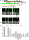
Functional characterization of monopartite and bipartite cNLSs in P. sojae transformants. (A) Subcellular localization of N-terminal cNLS-2XGFP fusions, involving mammalian cNLS sequences. SV40, c-Myc, NPL, represent cNLSs identified in SV40 large T antigen, human c-Myc proto-oncoprotein, and Xenopus nucleoplasmin respectively; H2B-mCherry = P. sojae histone H2B fused to the N-terminus of mCherry; Control = 2XGFP. “GFP” above the panel indicates visualization of the relevant GFP or 2xGFP fusions. Scale bar corresponds to 5 μm in this and all subsequent figures. (B) Subcellular localization of different fusions with the SV40 NLS. H2B-GFP was used as a positive control. (C) Exclusion of NPL-2XGFP from the nucleolus, as confirmed by P. capsici fibrillarin fused to mCherry (FIB-mCherry). (D) Quantification of nuclear localization by protein segments shown in (A,B). For quantification in all figures, LNC indicates the mean log2-transformed nuclear fluorescence to cytoplasmic fluorescence ratio from ~30 randomly selected pairs of nuclear and adjacent cytoplasmic regions. SV40 (C) = SV40 NLS attached at the C-terminus of 2XGFP (i.e., 2XGFP-SV40 NLS). Error bars, S.E. Asterisks, LNC-values of NLS-GFP fusions that are significantly greater than the LNC of 2XGFP alone (FDR p′ < 0.001). (E) Sequence alignment of predicted bipartite cNLSs tested in P. sojae. The proposed P. sojae bipartite cNLS consensus is shown on the top; [K/R] in red indicates the position of additional positive residues required in P. sojae. In the sequences of each protein, the two elements of each bipartite cNLS predicted by PSORT II (PSORT, 1997) are underlined, and basic amino acids within each cluster are in bold. The additional basic amino acids demonstrated to contribute to nuclear localization are in bold red. The residues in lowercase indicate non-native residues flanking the candidate NLS in each construct. F, functional NLS; NF, non-functional NLS. For PHYSO_561151 and PsL28, both the full length functional cNLSs and the truncated or mutant non-functional cNLSs are shown. (F,G) Functional tests of P. sojae bipartite NLSs listed in E. F, representative images. G, quantification. (1–33) m indicates mutation of the red-highlighted residues in PsL28 (K7A/R9A/K10A). Representative images are shown in (A–C,F).
Figure 2
Figure 2
PY-NLS prototypes exhibit weak nuclear targeting activities in P. sojae transformants. (A) Sequences of four well-characterized PY-NLSs derived from human or yeast proteins and the Kapβ2-specific nuclear import inhibitor, M9M peptide. Core residues that determine hydrophobic or basic PY-NLS type are shaded in yellow. The R/K/H-X2−5-PY/L consensus residues are bold and underlined. Residues marked in red and blue in M9M are sequences originating from M9 and hnRNP M respectively. (B) Subcellular localization of the five PY-NLS described in (A). Representative images are shown. (C) Quantification of localization of fusions observed in (B). For ease of comparison, the LNC-values of SV40 NLS-2XGFP and 2XGFP alone (from Figure 1) are included in the bar chart here and in subsequent figures; i.e., the same values were used in every figure. Asterisks, LNC-values of NLS-2XGFP fusions that are significantly greater (FDR p′ < 0.001) than the LNC of 2XGFP.
Figure 3
Figure 3
Subcellular localization produced by five protein segments containing candidate PY-NLS motifs. (A) The modified consensus sequence (Süel et al., 2008) used for searching for candidate PY-NLSs in P. sojae. Φ1 represents a hydrophobic residue (defined here as L, M, I, V, W, Y, H, A, P, or F) while Φ3 and Φ4 represent either hydrophobic residues or R or K./indicates alternative residues. Xm−n indicates any number of unspecified residues from m to n in number. (B) Nuclear localization of the full-length proteins PHYSO_357835, PHYSO_480605, PHYSO_251824, PHYSO_561151, and PHYSO_533817 in P. sojae transformants. Representative images. (C) Subcellular localization of PY-NLS-containing segments in P. sojae (left) and in HEK 293 cells (right). (D) HEK 293 cells expressing 2XGFP alone, SV40 NLS-2XGFP, and M9NLS-2XGFP, produced in the same experiment as controls. Representative images are shown in (B–D).
Figure 4
Figure 4
Nuclear localization of PHYSO_357835 is mediated by a PY-NLS that incorporates a cNLS-like motif. (A) Domain structure of PHYSO_357835. The position of the PY-NLS and the predicted cNLS within PHYSO_357835 are indicated by a black and a gray rectangle, respectively, and the corresponding amino acid sequence is listed below. The three PY-NLS epitopes are in bold, underlined, and numbered. Blue residues indicate the PY motif and basic region corresponding to the predicted cNLS, which were subjected to mutational analysis. No NLS sequences were predicted by NLStradamus or cNLS Mapper. (B) Subcellular localization of PHYSO_357835 mutants in the context of the C-terminal domain, 338–387. Left, representative images from P. sojae transformants expressing various mutations of 2XGFP-PHYSO_357835338−387. Right, quantification of the localization of 2XGFP-PHYSO_357835338−387 fusion proteins. (C) Subcellular localization of PHYSO_357835 mutants in the context of full length PHYSO_357835-GFP. Left, representative images; right, quantitation. The dots observed in P. sojae hyphae expressing PHYSO_357835-GFP-(R370A/H371A/K372A/R373A/P376A/Y377A) may be nucleoli as the WT shows substantial nucleolar localization, but this was not verified. The LNC for this mutant was calculated assuming the dots were nucleoli. Asterisks, LNC-values of NLS-2XGFP fusions that are significantly greater (FDR p′ < 0.001) than the LNC of 2XGFP.
Figure 5
Figure 5
Nuclear localization of PHYSO_480605 requires a region containing a PY-NLS with a variant PY motif. (A) Domain structure of PHYSO_480605. Position of the predicted PY-NLS is indicated by a black rectangle and the corresponding sequence is listed below. The three PY-NLS epitopes are in bold and underlined. The basic patch corresponding to a predicted cNLS is in bold and italics. No NLS sequences were predicted by PSORT II, NLStradamus or cNLS Mapper. (B) Subcellular localization of 2XGFP with full length PHYSO_480605 or fragments of it in P. sojae transformants. Left, representative images; right, quantification. Asterisks, LNC-values of NLS-(2X)GFP fusions that are significantly greater (FDR p′ < 0.001) than the LNC 2XGFP.
Figure 6
Figure 6
Nuclear localization of PHYSO_251824 requires contributions from two PY-NLS and one cNLS clustered within the C-terminus. (A) Domain structure of PHYSO_251824. The two candidate PY-NLSs and the two PSORT II-predicted cNLSs are indicated by black and gray rectangles respectively; their corresponding sequences are shown below. Predicted cNLS1 is inactive. The three epitopes of the two PY-NLS sequences are in bold and underlined. Amino acids subjected to mutational analysis are in blue. Amino acid sequences underlined by gray solid, dash, and dotted lines indicate the NLSs predicted by PSORT II, NLStradamus, and cNLS Mapper respectively. (B) Subcellular localization of PHYSO_251824-GFP and mutants fused to 2XGFP in P. sojae transformants. N-terminal truncations and cNLS2 were fused to 2XGFP at their C-termini, while various C-terminal PHYSO_251824 truncations were fused to 2XGFP at their N-termini. Representative images are shown. (C) Quantification of localization of PHYSO_251824 mutants. Image of PHYSO_251824239−358 is shown in Figure 3C. Mutational statuses of the various NLS candidates are labeled at the top. +, NLS candidate is the only one in the segment; -, NLS candidate is partially mutated; - -, NLS candidate is completely mutated. Asterisks, LNC-values of NLS-2XGFP fusions that are significantly greater (FDR p′ < 0.001) than the LNC of 2XGFP.
Figure 7
Figure 7
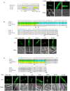
Combinatorial usage of NLSs for nuclear transport of P. sojae ribosomal protein L28 and core histones H3 and H4. (A–C) Upper panels, alignments of P. sojae ribosomal proteins L28 (PsL28, PHYSO_355737) and core histones H3 (PsH3, PHYSO_286415) and H4 (PsH4, PHYSO_285922) with their Arabidopsis thaliana (At), human (Hs), and Saccharomyces cerevisiae (Sc) orthologs. Asterisks on the top of each alignment indicate conserved residues among L28, H3, or H4 orthologs. Regions individually conferring nuclear targeting in human core histones are highlighted in green, cyan, or gray while those required in yeast are highlighted in yellow. NLSs predicted in the P. sojae core histone and ribosomal protein by PSORT II, NLStradamus, and NLS Mapper are underlined by gray solid, dash, and dotted lines respectively. Lower panels, subcellular localization of various PsL28, PsH3, or PsH4 truncations. Representative images are shown.

References

    1. Ah-Fong A. M., Judelson H. S. (2011). Vectors for fluorescent protein tagging in Phytophthora: tools for functional genomics and cell biology. Fungal Biol. 115, 882–890. 10.1016/j.funbio.2011.07.001 - DOI - PubMed
    1. Baake M., Bauerle M., Doenecke D., Albig W. (2001). Core histones and linker histones are imported into the nucleus by different pathways. Eur. J. Cell Biol. 80, 669–677. 10.1078/0171-9335-00208 - DOI - PubMed
    1. Benjamini Y., Hochberg Y. (1995). Controlling the false discovery rate - a practical and powerful approach to multiple testing. J. R. Statist. Soc. Ser. B Methodol. 57, 289–300.
    1. Bonifaci N., Moroianu J., Radu A., Blobel G. (1997). Karyopherin β2 mediates nuclear import of a mRNA binding protein. Proc. Natl. Acad. Sci. U.S.A. 94, 5055–5060. 10.1073/pnas.94.10.5055 - DOI - PMC - PubMed
    1. Cansizoglu A. E., Lee B. J., Zhang Z. C., Fontoura B. M., Chook Y. M. (2007). Structure-based design of a pathway-specific nuclear import inhibitor. Nat. Struct. Mol. Biol. 14, 452–454. 10.1038/nsmb1229 - DOI - PMC - PubMed

LinkOut - more resources