. 2016 Dec;12(4):318-327.
doi: 10.1183/20734735.012716.

Physiology of respiratory disturbances in muscular dystrophies

Affiliations

Physiology of respiratory disturbances in muscular dystrophies

Antonella Lo Mauro et al. Breathe (Sheff). 2016 Dec.

Abstract

Muscular dystrophy is a group of inherited myopathies characterised by progressive skeletal muscle wasting, including of the respiratory muscles. Respiratory failure, i.e. when the respiratory system fails in its gas exchange functions, is a common feature in muscular dystrophy, being the main cause of death, and it is a consequence of lung failure, pump failure or a combination of the two. The former is due to recurrent aspiration, the latter to progressive weakness of respiratory muscles and an increase in the load against which they must contract. In fact, both the resistive and elastic components of the work of breathing increase due to airway obstruction and chest wall and lung stiffening, respectively. The respiratory disturbances in muscular dystrophy are restrictive pulmonary function, hypoventilation, altered thoracoabdominal pattern, hypercapnia, dyspnoea, impaired regulation of breathing, inefficient cough and sleep disordered breathing. They can be present at different rates according to the type of muscular dystrophy and its progression, leading to different onset of each symptom, prognosis and degree of respiratory involvement.

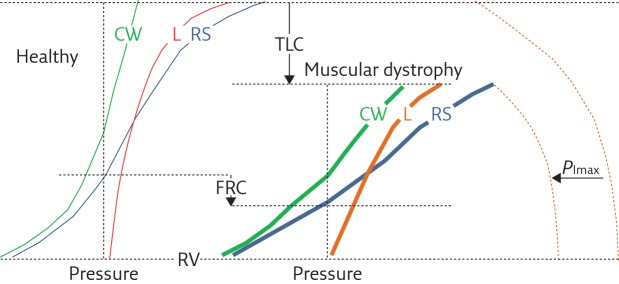
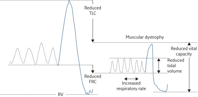
Key points: A common feature of muscular dystrophy is respiratory failure, i.e. the inability of the respiratory system to provide proper oxygenation and carbon dioxide elimination.In the lung, respiratory failure is caused by recurrent aspiration, and leads to hypoxaemia and hypercarbia.Ventilatory failure in muscular dystrophy is caused by increased respiratory load and respiratory muscles weakness.Respiratory load increases in muscular dystrophy because scoliosis makes chest wall compliance decrease, atelectasis and fibrosis make lung compliance decrease, and airway obstruction makes airway resistance increase.The consequences of respiratory pump failure are restrictive pulmonary function, hypoventilation, altered thoracoabdominal pattern, hypercapnia, dyspnoea, impaired regulation of breathing, inefficient cough and sleep disordered breathing.

Educational aims: To understand the mechanisms leading to respiratory disturbances in patients with muscular dystrophy.To understand the impact of respiratory disturbances in patients with muscular dystrophy.To provide a brief description of the main forms of muscular dystrophy with their respiratory implications.


Conflict of interest statement

Conflict of interest None declared.

Figures

Figure 1
Figure 1
Representative, schematic diagram showing the pressure–volume curves of the respiratory system (RS, blue curves) and its two components, i.e. the chest wall (CW, green curves) and lung (L, red curves), for healthy subjects (thin curves on the left panel) and patients with muscular dystrophy (thick curves on the right panel). The maximal inspiratory pressure (PImax) is also reported (dashed curves). Muscular dystrophy patients are characterised by 1) reduced total lung capacity (TLC); 2) reduced compliance of chest wall (CCW), lungs (CL) and respiratory system (slope of the corresponding pressure–volume curves); 3) reduced PImax; 4) reduced inspiratory capacity (IC=TLC−FRC); and 5) reduced expiratory reserve volume (ERV=FRC−RV). Functional residual capacity (FRC) may be lower or even normal. Residual volume (RV) is not significantly different than in healthy subjects. PImax represents the force of respiratory muscles, while the volume variations are the resulting action of their contraction. The fact that PImax and lung volumes are both reduced indicates that the respiratory muscles, and therefore the respiratory pump, are affected by muscular dystrophy. The compliance of the respiratory system reduces because it decreases in both its components: 1) CL, due to lung atelectasis and fibrosis, the former being a consequence of hypoventilation, induced by respiratory muscles involvement, the latter of recurrent aspiration, induced by the involvement of bulbar muscles; and 2) CCW, due to of the onset of scoliosis, induced by the involvement of trunk muscles.
Figure 2
Figure 2
Lung volume variations during quiet breathing (grey lines) and vital capacity (blue lines) in an healthy subject (left) and a muscular dystrophy patient (right). Muscular dystrophy patients are characterised by reduced vital capacity, and rapid and shallow breathing at rest, i.e. reduced tidal volume and increased respiratory rate. RV: residual volume; FRC: functional residual capacity.
Figure 3
Figure 3
Schematic diagram summarising the generalised causes of respiratory disturbances in muscular dystrophies. Raw: airway resistance; WOBEL: elastic component of work of breathing; WOBRES: resistive component of work of breathing; ↑: increment; ↓: decrement.
Figure 4
Figure 4
a) Ribcage (top), abdominal (middle) and chest wall (bottom) volume variations during spontaneous breathing in supine position in a 17-year-old DMD patient. While tidal volume is constant, the thoracoabdominal pattern alternatively changes by passing from abdominal (A, blue lines) to thoracic (B, grey lines) predominance. b) “Loops” (i.e. volume changes of the abdomen versus the ribcage) in representative breaths when either the abdomen (A) or the ribcage (B) are prevalent. The breath of a healthy peer (control) is also reported. The arrows indicate the direction of the loops, i.e. the relative action of inspiratory ribcage muscles and the diaphragm [31]. In healthy subjects, both ribcage and abdomen expand during inspiration, with the latter being predominant. This indicates that in the supine position, the diaphragm is the leading muscle of inspiration, with a reduced contribution of ribcage muscles that mainly act to avoid chest wall distortion. The DMD patient therefore alternates periods of breathing in which inspiration is led by the diaphragm (A) and periods in which ribcage muscles are the leading inspiratory muscles (B). These thoracoabdominal “alternans” are thought to be a strategy adopted to cope with diaphragmatic weakness and fatigue. Note that, however, when the diaphragm leads inspiration (A), its action is reduced compared to the healthy subject (i.e. reduced abdominal expansion) and this is an index of diaphragmatic weakness.

References

    1. Emery AEH. The muscular dystrophies. Lancet 2002; 359: 687–695. - PubMed
    1. Perrin C, Unterborn JN, Ambrosio CD, et al. Pulmonary complications of chronic neuromuscular diseases and their management. Muscle Nerve 2004; 29: 5–27. - PubMed
    1. Allen J. Pulmonary complications of neuromuscular disease: A Respiratory mechanics perspective. Paediatr Respir Rev 2010; 11: 18–23. - PubMed
    1. De Troyer A. Respiratory effect of the lower rib displacement produced by the diaphragm. J Appl Physiol 2012; 112: 529–534. - PubMed
    1. Criner GJ, Marchetti N. Effects of neuromuscular diseases on ventilation. In: Grippi MA, Elias JA, Fishman JA, et al. eds. Fishman’s Pulmonary Diseases and Disorders. 5th Edn New York, McGraw-Hill Medical, 2015; pp. 1289–1311.