

Zygomatic Intraosseous Hemangioma: Case Report and Literature Review
- PMID: 28210401
- PMCID: PMC5309129
- DOI: 10.1055/s-0036-1592087
Zygomatic Intraosseous Hemangioma: Case Report and Literature Review
Abstract
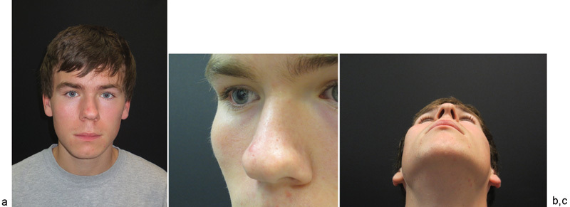
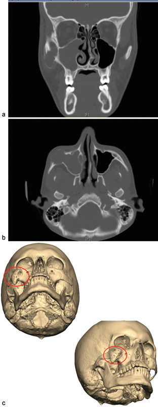
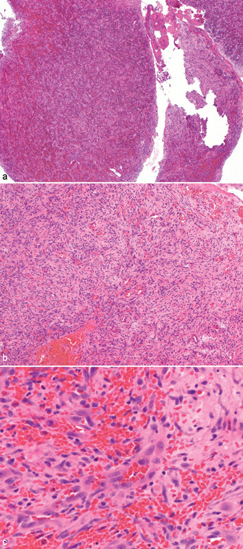
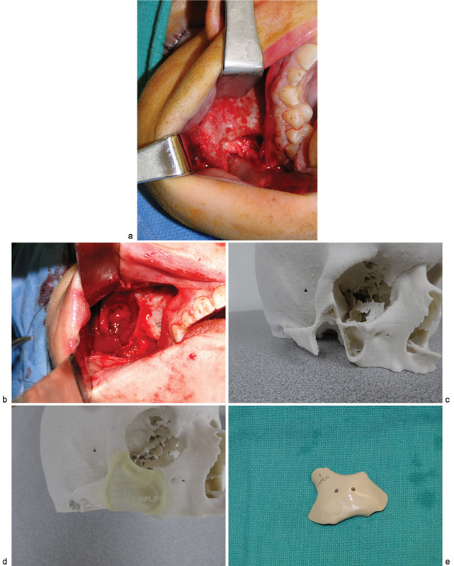
Intraosseous hemangiomas are uncommon intrabony lesions, representing approximately 0.5 to 1% of all intraosseous tumors. Their description varies from "benign vasoformative neoplasms" to true hamartomatous proliferations of endothelial cells forming a vascular network with intermixed fibrous connective tissue stroma. These commonly present as a firm, painless swelling. Intraosseous hemangiomas present more commonly in females than in males and most likely occur in the fourth decade of life. The most common etiology of intraosseous hemangioma is believed to be prior trauma to the area. They have a tendency to bleed briskly upon removal or biopsy, making preoperative detection of the vascular nature of the lesion of significant importance. There are four variants: (1) capillary type, (2) cavernous type, (3) mixed variant, and (4) scirrhous type. Generally most common in the vertebral skeleton, they can also present in the calvarium and facial bones. In the head, the most common site is the parietal bone, followed by the mandible, and then malar and zygomatic regions. Intraosseous hemangiomas of the zygoma are rare entities with the first case reported in 1950 by Schoenfield. In this article, we review 49 case reports of intraosseous hemangioma of the zygoma, and also present a new case treated with excision followed by polyether-ether ketone implant placement for primary reconstruction.
Keywords: PEEK implant; intraosseous hemangioma; zygoma.
Figures
References
-
- Marshak G. Hemangioma of the zygomatic bone. Arch Otolaryngol. 1980;106(9):581–582. - PubMed
-
- Ethunandan M, Mellor T K. Haemangiomas and vascular malformations of the maxillofacial region—a review. Br J Oral Maxillofac Surg. 2006;44(4):263–272. - PubMed
-
- Zucker J J, Levine M R, Chu A. Primary intraosseous hemangioma of the orbit. Report of a case and review of literature. Ophthal Plast Reconstr Surg. 1989;5(4):247–255. - PubMed
Publication types
LinkOut - more resources
Full Text Sources
Other Literature Sources