

Eye Inside Out: Endonasal Endoscopic Reposition of Eye from Nose with Complete Vision Regainment
- PMID: 28210414
- PMCID: PMC5305312
- DOI: 10.1055/s-0036-1584401
Eye Inside Out: Endonasal Endoscopic Reposition of Eye from Nose with Complete Vision Regainment
Abstract
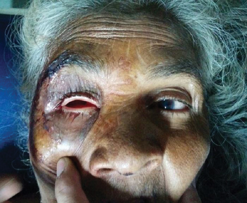
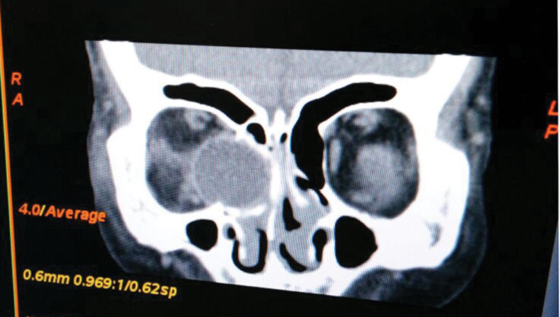
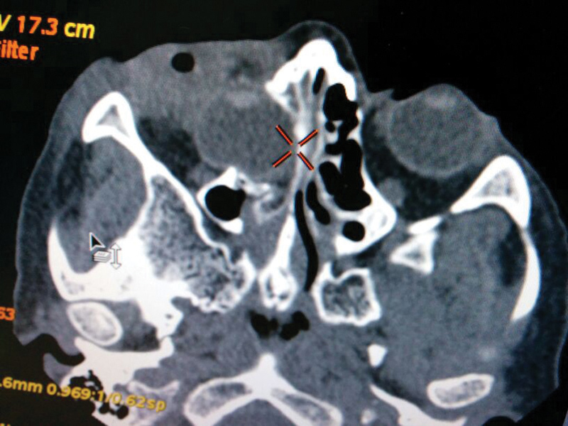
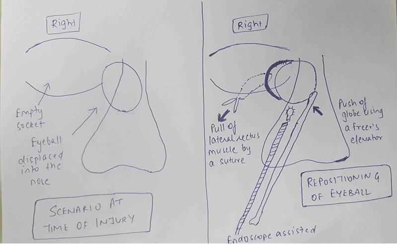
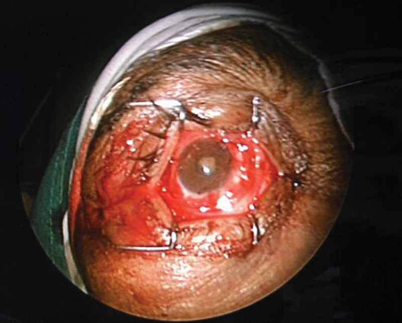
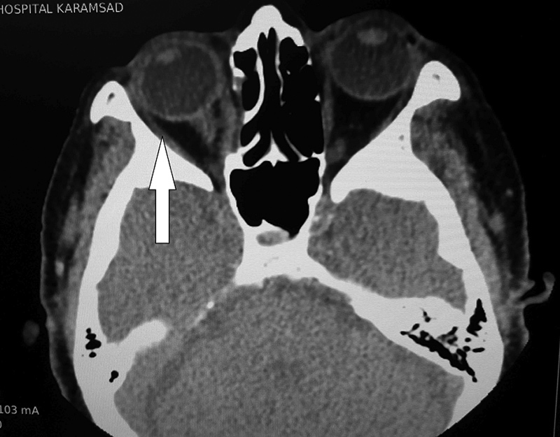
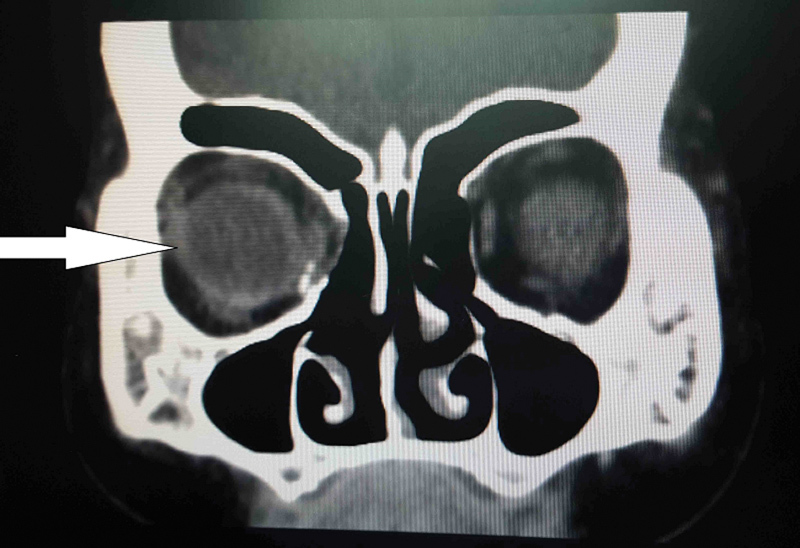
Faciomaxillary and ocular trauma is a common entity in most emergency and trauma units. We came across a 68-year-old female patient with a history of bull horn injury over the right eye. Examination revealed an empty orbital socket with unreliable perception of light present. Imaging showed that the eye had displaced posteroinferomedially to be lying in the ethmoid air cells in the nasal cavity. Under nasal endoscopic guidance, the eye was reposited back into the orbital socket and conjunctival sutures were taken to stabilize the position. The patient had vision of counting fingers at 1.5 m on the first postoperative day which improved to 6/24 on last follow-up. Such is the rarity that never before has such a case been described in literature where traumatic displacement of eyeball into the nose has been successfully repositioned by an endonasal endoscope with appreciable regaining of vision. It also further promotes endonasal endoscopic approach in the management of orbital blow out injuries.
Keywords: bull horn injury; endonasal endoscopic repair; medial orbital blow out; ocular trauma.
Figures

Similar articles
-
Traumatic eye ball luxation: A stepwise approach to globe salvage.Saudi J Ophthalmol. 2017 Oct-Dec;31(4):260-265. doi: 10.1016/j.sjopt.2017.06.001. Epub 2017 Jun 13. Saudi J Ophthalmol. 2017. PMID: 29234230 Free PMC article.
-
[The treatment of nose-eye correlated diseases with external nasal incision combined with nasal cavity approach surgery through endoscope].Lin Chuang Er Bi Yan Hou Tou Jing Wai Ke Za Zhi. 2016 Aug 5;30(15):1210-1214. doi: 10.13201/j.issn.1001-1781.2016.15.008. Lin Chuang Er Bi Yan Hou Tou Jing Wai Ke Za Zhi. 2016. PMID: 29798331 Chinese.
-
Overcorrection of a Medial Orbital Wall Fracture Using the Endonasal Approach.J Craniofac Surg. 2016 Oct;27(7):1837-1838. doi: 10.1097/SCS.0000000000002944. J Craniofac Surg. 2016. PMID: 27483095
-
Endoscopic endonasal optic nerve and orbital apex decompression for nontraumatic optic neuropathy: surgical nuances and review of the literature.Neurosurg Focus. 2014;37(4):E19. doi: 10.3171/2014.7.FOCUS14303. Neurosurg Focus. 2014. PMID: 25270138 Review.
-
Tension pneumo-orbit treated by endoscopic, endonasal decompression: case report and literature review.J Laryngol Otol. 2008 Mar;122(3):e8. doi: 10.1017/S002221510700165X. Epub 2008 Feb 11. J Laryngol Otol. 2008. PMID: 18267045 Review.
References
-
- Ko M J, Morris C K, Kim J W, Lad S P, Arrigo R T, Lad E M. Orbital fractures: national inpatient trends and complications. Ophthal Plast Reconstr Surg. 2013;29(4):298–303. - PubMed
-
- Nirmalan P K, Katz J, Tielsch J M. et al.Ocular trauma in a rural south Indian population: the Aravind Comprehensive Eye Survey. Ophthalmology. 2004;111(9):1778–1781. - PubMed
-
- Burm J S, Chung C H, Oh S J. Pure orbital blowout fracture: new concepts and importance of medial orbital blowout fracture. Plast Reconstr Surg. 1999;103(7):1839–1849. - PubMed
-
- Motamedi M H, Dadgar E, Ebrahimi A, Shirani G, Haghighat A, Jamalpour M R. Pattern of maxillofacial fractures: a 5-year analysis of 8,818 patients. J Trauma Acute Care Surg. 2014;77(4):630–634. - PubMed
Publication types
LinkOut - more resources
Full Text Sources
Other Literature Sources