

Unmasking histoplasmosis immune reconstitution inflammatory syndrome in a patient recently started on antiretroviral therapy
- PMID: 28210571
- PMCID: PMC5304559
- DOI: 10.4322/acr.2016.048
Unmasking histoplasmosis immune reconstitution inflammatory syndrome in a patient recently started on antiretroviral therapy
Erratum in
-
Erratum.Autops Case Rep. 2017 Mar 30;7(1):55. doi: 10.4322/acr.2017.011. eCollection 2017 Jan-Mar. Autops Case Rep. 2017. PMID: 28536689 Free PMC article.
Abstract
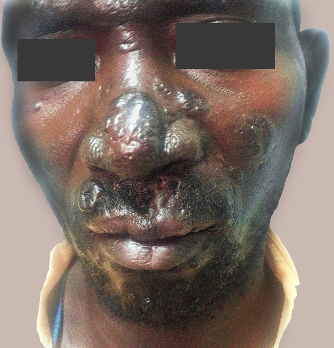
Histoplasmosis is the most common endemic mycoses among HIV-infected people. Patients with suppressed cell immunity mainly due to HIV are at increased risk of disseminated disease. Dermatological manifestations of immune reconstitution inflammatory syndrome (IRIS) and cutaneous manifestations of histoplasmosis similar to an IRIS event have been previously described. We report the case of a 43-year-old male who presented with cutaneous disseminated histoplasmosis due to Histoplasma capsulatum var. capsulatum 4 months after the onset of the antiretroviral therapy and some improvement in the immune reconstitution. After 2 weeks of amphotericin B and itraconazole therapy, the scheduled treatment involved fluconazole maintenance therapy, which resulted in an improvement of his skin lesions.
Keywords: Antiretroviral Therapy, Highly Active, Fluconazole; Histoplasmosis; Immune Reconstitution Inflammatory Syndrome.
Conflict of interest statement
Conflict of interest: None
Figures


Similar articles
-
Incidence and Trends in Immune Reconstitution Inflammatory Syndrome Associated With Histoplasma capsulatum Among People Living With Human Immunodeficiency Virus: A 20-Year Case Series and Literature Review.Clin Infect Dis. 2020 Feb 3;70(4):643-652. doi: 10.1093/cid/ciz247. Clin Infect Dis. 2020. PMID: 30921453 Review.
-
Histoplasmosis-associated immune reconstitution inflammatory syndrome.An Bras Dermatol. 2011 Jul-Aug;86(4 Suppl 1):S168-72. doi: 10.1590/s0365-05962011000700044. An Bras Dermatol. 2011. PMID: 22068802 English, Portuguese.
-
Disseminated cutaneous histoplasmosis with laryngeal involvement in a setting of immune reconstitution inflammatory syndrome.South Afr J HIV Med. 2017 Apr 28;18(1):693. doi: 10.4102/sajhivmed.v18i1.693. eCollection 2017. South Afr J HIV Med. 2017. PMID: 29568629 Free PMC article.
-
[African histoplasmosis due to Histoplasma capsulatum var. duboisii: relationship with AIDS in recent Congolese cases].Sante. 1995 Jul-Aug;5(4):227-34. Sante. 1995. PMID: 7582643 French.
-
Disseminated histoplasmosis caused by Histoplasma capsulatum var. duboisii in a non-HIV patient in Burkina Faso: Case report.J Mycol Med. 2015 Jun;25(2):159-62. doi: 10.1016/j.mycmed.2015.03.002. Epub 2015 Mar 31. J Mycol Med. 2015. PMID: 25840849 Review.
Cited by
-
Histoplasma stomatitis unveiled: Not all opportunistic infections get better after initiation of antiretroviral therapy.Clin Case Rep. 2021 Jan 19;9(3):1466-1468. doi: 10.1002/ccr3.3799. eCollection 2021 Mar. Clin Case Rep. 2021. PMID: 33768869 Free PMC article.
-
Opportunistic Invasive Mycoses in AIDS: Cryptococcosis, Histoplasmosis, Coccidiodomycosis, and Talaromycosis.Curr Infect Dis Rep. 2017 Aug 22;19(10):36. doi: 10.1007/s11908-017-0592-7. Curr Infect Dis Rep. 2017. PMID: 28831671 Review.
-
Understanding Pathogenesis and Care Challenges of Immune Reconstitution Inflammatory Syndrome in Fungal Infections.J Fungi (Basel). 2018 Dec 17;4(4):139. doi: 10.3390/jof4040139. J Fungi (Basel). 2018. PMID: 30562960 Free PMC article. Review.
-
Left hand extensor tenosynovitis due to Histoplasma capsulatum complicated by immune reconstitution inflammatory syndrome.J Bone Jt Infect. 2021 Sep 23;6(8):355-361. doi: 10.5194/jbji-6-355-2021. eCollection 2021. J Bone Jt Infect. 2021. PMID: 34611507 Free PMC article.
-
Re-estimation of the burden of serious fungal diseases in Uganda.Ther Adv Infect Dis. 2024 Feb 6;11:20499361241228345. doi: 10.1177/20499361241228345. eCollection 2024 Jan-Dec. Ther Adv Infect Dis. 2024. PMID: 38328511 Free PMC article.
References
-
- Wheat LJ. Histoplasmosis: a review for clinicians from non-endemic areas. Mycoses. 2006;49(4):274-82. http://dx.doi.org/10.1111/j.1439-0507.2006.01253.x. PMid:. - DOI - PubMed
-
- Cottle LE, Gkrania-Klotsas E, Williams HJ, et al. . A multinational outbreak of histoplasmosis following a biology field trip in the Ugandan rainforest. J Travel Med. 2013;20(2):83-7. http://dx.doi.org/10.1111/jtm.12012. PMid:. - DOI - PubMed
-
- Kurowski R, Ostapchuk M. Overview of histoplasmosis. Am Fam Physician. 2002;66(12):2247-52. PMid:. - PubMed
-
- Kauffman CA. Histoplasmosis: a clinical and laboratory update. Clin Microbiol Rev. 2007;20(1):115-32. http://dx.doi.org/10.1128/CMR.00027-06. PMid:. - DOI - PMC - PubMed
Publication types
LinkOut - more resources
Full Text Sources
Other Literature Sources
