. 2015 Jan 3;1(2):203-221.e3.
doi: 10.1016/j.jcmgh.2014.12.003. eCollection 2015 Mar.

IL33 Is a Stomach Alarmin That Initiates a Skewed Th2 Response to Injury and Infection

Affiliations

IL33 Is a Stomach Alarmin That Initiates a Skewed Th2 Response to Injury and Infection

Jon N Buzzelli et al. Cell Mol Gastroenterol Hepatol. .

Abstract

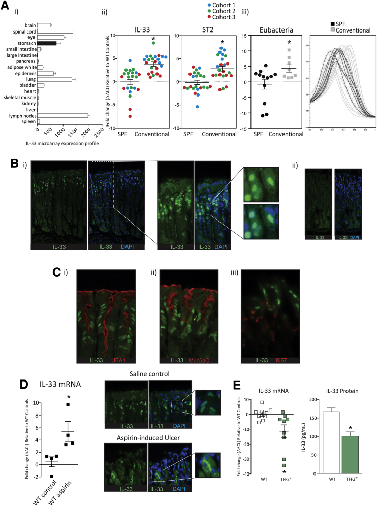
Background & aims: Interleukin (IL)33 is a recently described alarmin that is highly expressed in the gastric mucosa and potently activates Th2 immunity. It may play a pivotal role during Helicobacter pylori infection. Here, we delineate the role of IL33 in the normal gastric mucosa and in response to gastropathy.

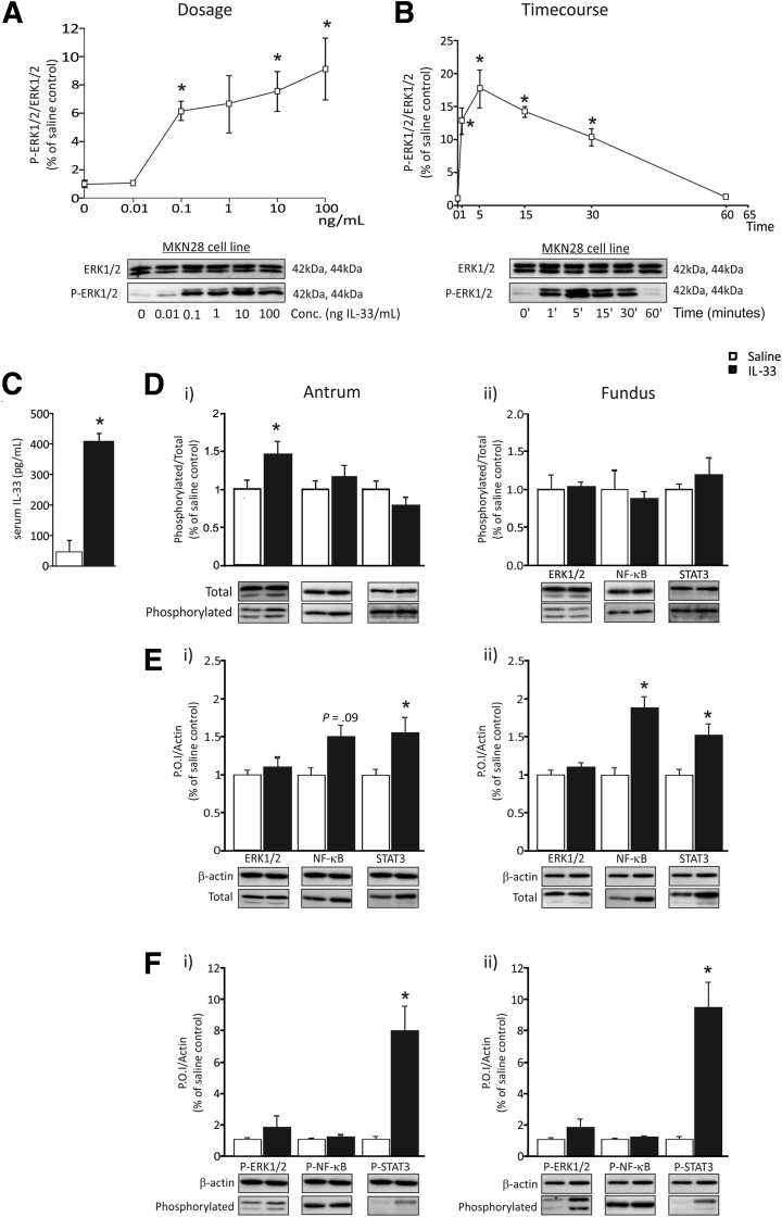
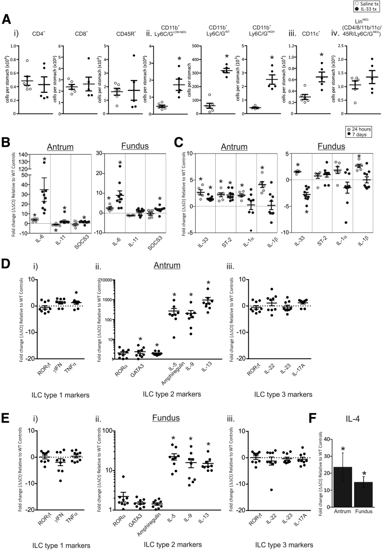
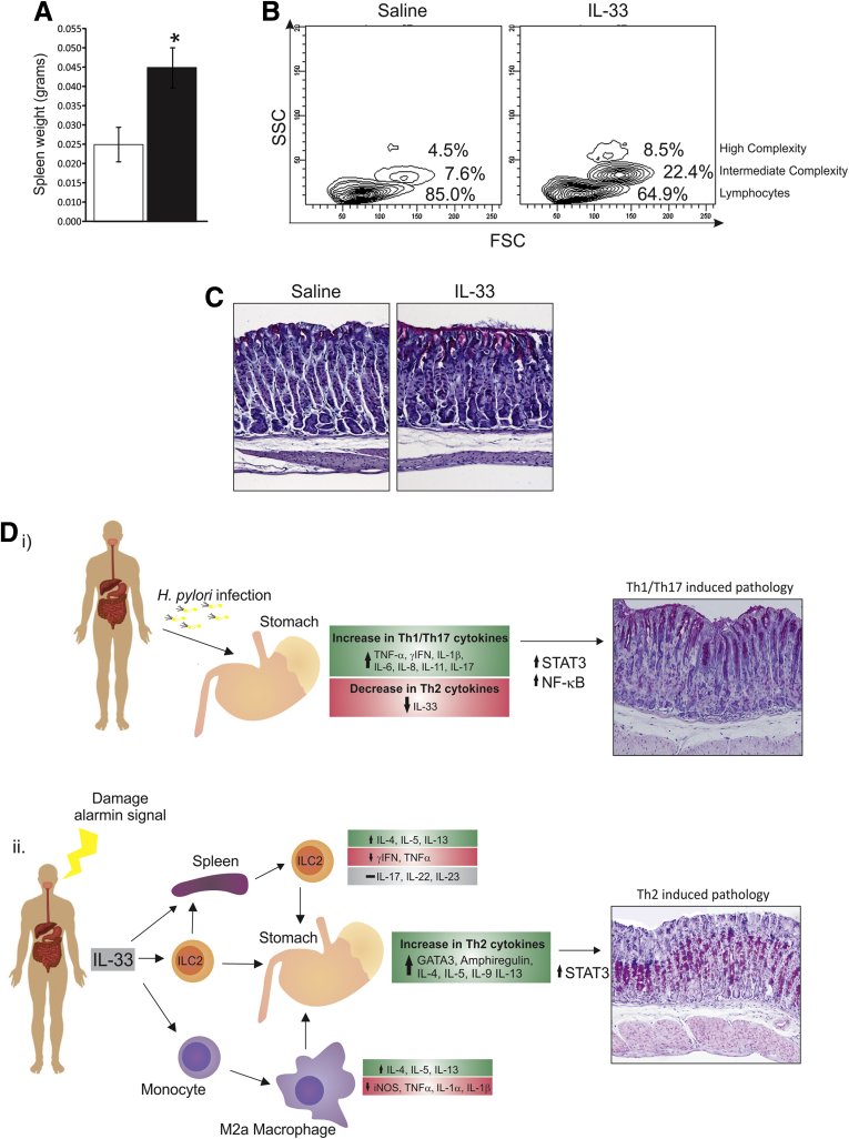
Methods: IL33 expression was evaluated in mice and human biopsy specimens infected with H pylori and in mice after dosing with aspirin. IL33 expression was localized in the gastric mucosa using immunofluorescence. Mice were given 1 or 7 daily doses of recombinant IL33 (1 μg/dose), and the stomach and the spleen responses were quantified morphologically, by flow cytometry and using quantitative reverse-transcription polymerase chain reaction and immunoblotting.

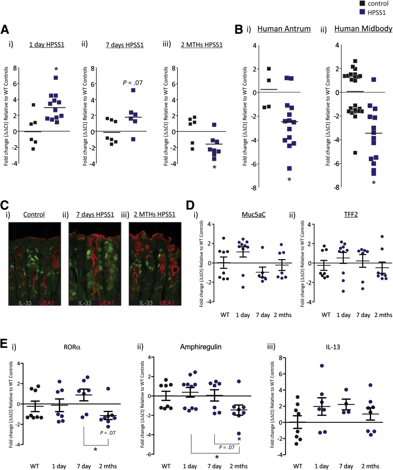
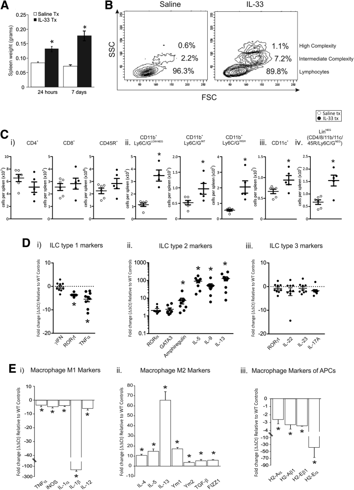
Results: In mice, the IL33 protein was localized to the nucleus of a subpopulation of surface mucus cells, and co-localized with the surface mucus cell markers Ulex Europaeus 1 (UEA1), and Mucin 5AC (Muc5AC). A small proportion of IL33-positive epithelial cells also were Ki-67 positive. IL33 and its receptor Interleukin 1 receptor-like 1 (ST2) were increased 4-fold after acute (1-day) H pylori infection, however, this increase was not apparent after 7 days and IL33 expression was reduced 2-fold after 2 months. Similarly, human biopsy specimens positive for H pylori had a reduced IL33 expression. Chronic IL33 treatment in mice caused systemic activation of innate lymphoid cell 2 and polarization of macrophages to the M2 phenotype. In the stomach, IL33-treated mice developed transmural inflammation and mucous metaplasia that was mediated by Th2/signal transducer and activator of transcription 3 signaling. Rag-1-/- mice, lacking mature lymphocytes, were protected from IL33-induced gastric pathology.

Conclusions: IL33 is highly expressed in the gastric mucosa and promotes the activation of T helper 2-cytokine-expressing cells. The loss of IL33 expression after prolonged H pylori infection may be permissive for the T helper 1-biased immune response observed during H pylori infection and subsequent precancerous progression.

Keywords: AB, Alcian blue; DC, dendritic cell; ELISA, enzyme-linked immunosorbent assay; ERK, extracellular signal–regulated kinase; FBS, fetal bovine serum; Gastric Cancer; HBSS, Hank’s balanced salt solution; Helicobacter pylori; IL, interleukin; IL33; ILC, innate lymphoid cell; Inflammatory Response; NF-κB, nuclear factor-κB; PAS, periodic acid–Schiff; PCR, polymerase chain reaction; QRT-PCR, quantitative reverse-transcription polymerase chain reaction; SMC, surface mucus cells; SPF, specific pathogen free; SS1, Sydney strain 1; STAT, signal transducer and activator of transcription; TFF, trefoil factor; Th, T-helper; WT, wild type; mRNA, messenger RNA.


Figures

Figure 1
Figure 1
IL33 localization in the gastric mucosa and mRNA expression increase owing to bacterial load and gastric insult. (Ai) Comparison of IL33 expression in mouse tissues (repeated twice). (Aii) IL33 and ST2 mRNA expression in the fundus of mice housed in either SPF or conventional facilities as well as mouse gastric bacterial load (N ≥ 20) (repeated 3 times). (Aiii) Eubacterial 16S expression in the SPF and conventional animal facility. (Bi) IL33 protein localization in the gastric mucosa; (ii) IL33 immunofluorescence on the gastric mucosa of IL33-/- mice (N = 5) (repeated twice). (C) Co-localization of IL33 with (i) UEA1, (ii) Muc5AC, and (iii) Ki-67 in the gastric mucosa (N = 6). (D) IL33 mRNA 4 hours after aspirin insult and IL33 protein localization at gastric ulcers (repeated twice, N = 6). (E) IL33 mRNA and protein levels in 12-week-old WT and TFF2-/- mice measured by QRT-PCR and ELISA assay, respectively (N = 8) (repeated twice). ∗Statistically significant, P ≤ .05. DAPI, 4′,6-diamidino-2-phenylindole.
Figure 2
Figure 2
IL33 mRNA expression in the stomach increases after acute H pylori infection and steadily decreases with more chronic infection. (A) IL33 mRNA expression after H pylori SS1 infection in mice; (i) 1 day, (ii) 7 days, and (iii) 2 months relative to uninfected mice and standardized to the housekeeper L32 (N ≥ 6) (repeated twice). (B) IL33 mRNA expression in human biopsy specimens positive for H pylori infection relative to uninfected biopsy specimens and standardized to the housekeeper L32; (i) antrum and (ii) midbody (N ≥ 12). (C) Co-localization of IL33 with UEA1 in H pylori–SS1–infected mice: (i) control, (ii) 7 days, and (iii) 2 months (N = 5). (D) mRNA expression of (i) Muc5AC and (ii) TFF2 after 1 day, 7 days, and 2 months of H pylori–SS1 infection, and standardized to the housekeeper L32 (N ≥ 6). (E) Expression of the ILC2 genes: (i) RORα, (ii) amphiregulin, and (iii) IL13 after 1 day, 7 days, and 2 months of H pylori–SS1 infection, and standardized to the housekeeper L32 (N ≥ 6). ∗Statistically significant, P ≤ .05.
Figure 3
Figure 3
The gastric mucosa is responsive to IL33 both in vitro and in vivo. (A) Dose-dependent responsiveness of MKN28 cells to IL33 assessed through analysis of ERK1/2 activation (N = 3) (repeated twice). (B) Time-dependent responsiveness of MKN28 cells to IL33 assessed through analysis of ERK1/2 activation (N = 3) (repeated twice). (C) IL33 protein levels in sera of mice treated with IL33 for 7 days measured by ELISA. (D) Phosphorylation of ERK1/2, NF-κB, and STAT3 relative to total protein in the antrum and fundus of mice 24 hours after IL33 administration. (E) Amount of total protein of ERK1/2, NF-κB, and STAT3 relative to the housekeeping protein β-actin in the antrum and fundus of mice treated with IL33 for 7 days. (F) Amount of phosphorylation protein of ERK1/2, NF-κB, and STAT3 relative to the housekeeping protein β-actin in the antrum and fundus of mice treated with IL33 for 7 days. Saline, n = 10; IL33, n = 9; experimented was repeated 3 times. ∗Statistically significant, P ≤ .05. P.O.I., protein of interest.
Figure 4
Figure 4
IL33 treatment causes atypical gastric pathology. (A) AB-PAS comparison of the cardiac region of the stomach between saline- and IL33-treated mice. (B) AB-PAS comparison of the antrum between saline- and IL33-treated mice. (C) AB-PAS comparison of the fundus between saline- and IL33-treated mice, and semiquantitative assessment of gastric inflammation, atrophy, and metaplasia. (D) Assessment of parietal cell atrophy with immunohistochemistry using antibody directed to H+/K+ adenosine triphosphatase β subunit. (E) Assessment of proliferation in the fundic glands with immunohistochemistry using antibody directed to Ki-67. (F) Assessment of chief cell atrophy with immunohistochemistry using antibody directed to intrinsic factor. (G) Assessment of mast cell infiltrate in the fundus with immunohistochemistry using antibody directed to mast cell chymase and mRNA expression of mast cell markers in IL33-treated mice relative to saline-treated mice and standardized to the housekeeper L32. Saline, n = 10; IL33, n = 9; experiment was repeated 3 times. ∗Statistically significant, P ≤ .05. Tx, treatment.
Figure 5
Figure 5
Effects of IL33 on the spleen and peritoneal macrophages. (A) Spleen weight after 24 hours and 7 days of IL33 treatment. (B) Size and complexity of immune cells in the spleen after 7 days of IL33 treatment compared with saline controls. (C) Total number of immunocyte types in the spleens of IL33-treated mice compared with saline controls: (i) CD4+, CD8+, and CD45R+; (ii) CD11b+Ly6C/GLOW-NEG, CD11b+Ly6C/GINT, and CD11b+Ly6C/GHIGH; (iii) CD11c+; and (iv) lineage-negative immunocytes (saline, n = 6; IL33, n = 5). (D) mRNA expression of markers of ILC in the spleen of 7-day IL33-treated mice all expressed relative to saline-treated mice and standardized to the housekeeper L32: (i) group 1 ILC markers; (ii) group 2 ILC markers; and (iii) group 3 ILC markers. (E) Assessment of peritoneal macrophage mRNA expression profile after IL33 treatment all expressed relative to saline-treated mice and standardized to the housekeeper L32: (i) M1 markers, (ii) M2 markers, (iii) markers of antigen-presenting cells (APCs). Saline, n = 10; IL33, n = 9; experiment was repeated twice. ∗Statistically significant, P ≤ .05. FSC, forward scatter; iNOS, inducible nitric oxide synthase; SSC, side scatter; Tx, treatment.
Figure 6
Figure 6
Immunocyte populations in the stomach of 7-day IL33-treated mice and mRNA expression profile of ligands and regulators of gp130, IL1 cytokines, and ILC markers in the gastric mucosa of 24-hour and 7-day IL33-treated mice. (A) Total number of immunocyte types in the stomach of IL33-treated mice compared with saline controls: (i) CD4+, CD8+, and CD45R+; (ii) CD11b+Ly6C/GLOW-NEG, CD11b+Ly6C/GINT, and CD11b+Ly6C/GHIGH; (iii) CD11c+; and (iv) lineage-negative immunocytes (saline, n = 6; IL33, n = 5). (B) mRNA expression of gp130 ligands in the antrum and fundus of 24-hour and 7-day IL33-treated mice all expressed relative to saline-treated mice and standardized to the housekeeper L32. (C) mRNA expression of IL1 family cytokines in the antrum and fundus of 24-hour and 7-day IL33-treated mice. (D) Antrum and (E) fundus, expression of ILC markers: (i) group 1 ILC markers; (ii) group 2 ILC markers; and (iii) group 3 ILC markers. (E) IL4 expression in the antrum and fundus of 7-day IL33-treated mice. All values are expressed relative to saline-treated mice and standardized to the housekeeper L32. Saline, n = 10; IL33, n = 9; experiment repeated twice. ∗Statistically significant, P ≤ .05.
Figure 7
Figure 7
Effects of IL33 on the spleen and stomach of lymphocyte-deficient mice. (A) Spleen weight of IL33-treated Rag-1-/- mice. (B) Size and complexity of immune cells in the spleen of 7-day IL33-treated Rag-1-/- mice. (C) AB-PAS staining of the fundus of Rag-1-/- mice. WT-saline, n = 6; WT–IL33, n = 6; Rag-1-/-–saline, n = 6; Rag-1-/-–IL33, n = 6. (D) Summary of immunologic changes and gastric pathology after (i) H pylori infection or (ii) 7 days of IL33 treatment. ∗Statistically significant, P ≤ .05. SSC, standard saline citrate.

References

    1. Oppenheim J.J., Yang D. Alarmins: chemotactic activators of immune responses. Curr Opin Immunol. 2005;17:359–365. - PubMed
    1. Arshad M.I., Piquet-Pellorce C., Samson M. IL-33 and HMGB1 alarmins: sensors of cellular death and their involvement in liver pathology. Liver Int. 2012;32:1200–1210. - PubMed
    1. Schmitz J., Owyang A., Oldham E. IL-33, an interleukin-1-like cytokine that signals via the IL-1 receptor-related protein ST2 and induces T helper type 2-associated cytokines. Immunity. 2005;23:479–490. - PubMed
    1. Neill D.R., Wong S.H., Bellosi A. Nuocytes represent a new innate effector leukocyte that mediates type-2 immunity. Nature. 2010;464:1367–1370. - PMC - PubMed
    1. Pecaric-Petkovic T., Didichenko S.A., Kaempfer S. Human basophils and eosinophils are the direct target leukocytes of the novel IL-1 family member IL-33. Blood. 2009;113:1526–1534. - PMC - PubMed

LinkOut - more resources