. 2017 Mar;112(2):18.
doi: 10.1007/s00395-017-0606-5. Epub 2017 Feb 16.

Repeated doses of cardiac mesenchymal cells are therapeutically superior to a single dose in mice with old myocardial infarction

Affiliations

Repeated doses of cardiac mesenchymal cells are therapeutically superior to a single dose in mice with old myocardial infarction

Yiru Guo et al. Basic Res Cardiol. 2017 Mar.

Abstract

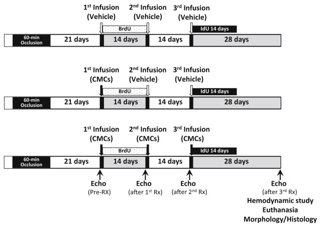
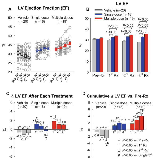
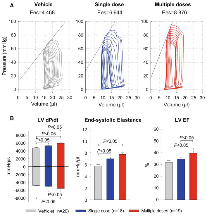
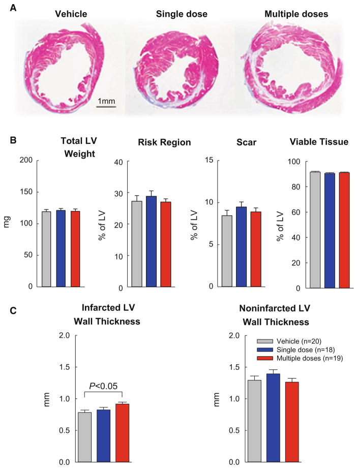
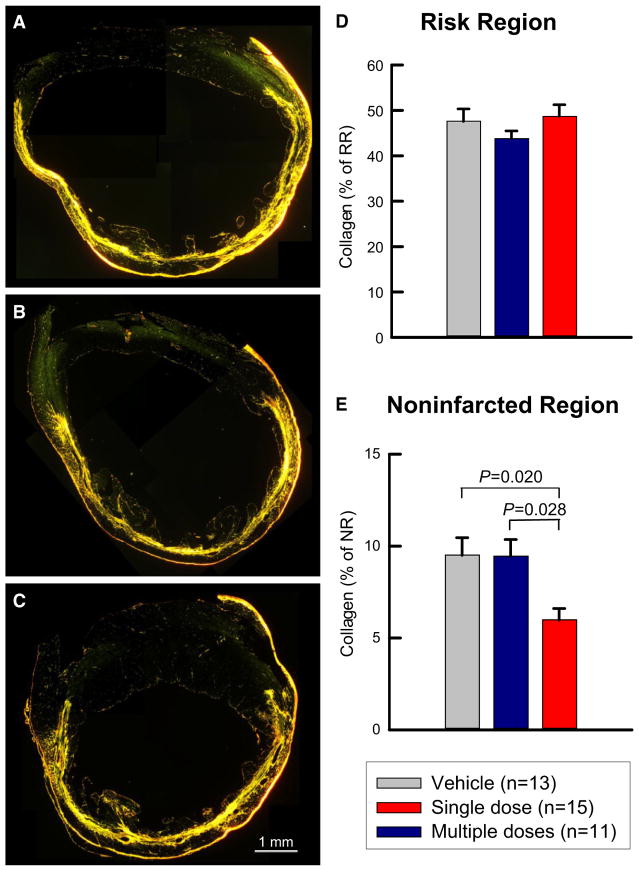
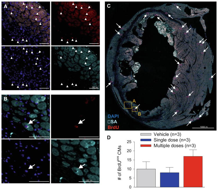
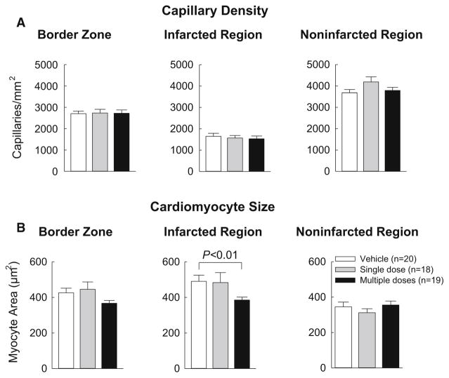
We have recently demonstrated that repeated administrations of c-kitPOS cardiac progenitor cells (CPCs) have cumulative beneficial effects in rats with old myocardial infarction (MI), resulting in markedly greater improvement in left ventricular (LV) function compared with a single administration. To determine whether this paradigm applies to other species and cell types, mice with a 3-week-old MI received one or three doses of cardiac mesenchymal cells (CMCs), a novel cell type that we have recently described. CMCs or vehicle were infused percutaneously into the LV cavity, 14 days apart. Compared with vehicle-treated mice, the single-dose group exhibited improved LV ejection fraction (EF) after the 1st infusion (consisting of CMCs) but not after the 2nd and 3rd (vehicle). In contrast, in the multiple-dose group, LV EF improved after each CMC infusion, so that at the end of the study, LV EF averaged 35.5 ± 0.7% vs. 32.7 ± 0.6% in the single-dose group (P < 0.05). The multiple-dose group also exhibited less collagen in the non-infarcted region vs. the single-dose group. Engraftment and differentiation of CMCs were negligible in both groups, indicating paracrine effects. These results demonstrate that, in mice with ischemic cardiomyopathy, the beneficial effects of three doses of CMCs are significantly greater than those of one dose, supporting the concept that multiple treatments are necessary to properly evaluate the full therapeutic potential of cell therapy. Thus, the repeated-treatment paradigm is not limited to c-kit POS CPCs or to rats, but applies to other cell types and species. The generalizability of this concept dramatically augments its significance.

Keywords: Cell therapy; Ischemic cardiomyopathy; Progenitor cells; Stem cells.


Figures

Fig. 1
Fig. 1
Experimental protocol. Mice were subjected to a 60-min coronary artery occlusion followed by reperfusion to induce myocardial infarction (MI). Three weeks after MI, animals received the 1st treatment (vehicle or 1 × 10⁶ CMCs) via echo-guided LV infusion, followed by a 2nd and 3rd treatment at 2-week intervals. BrdU was given right after the 1st treatment until the 2nd treatment; IdU was given for 2 weeks after the 3rd treatment
Fig. 2
Fig. 2
Myocardial retention of cells 24 h after injection. Mice underwent a 60-min coronary occlusion; three weeks later, 1×10⁶ CMCs were injected percutaneously into the LV cavity under echo guidance. 5 min and 24 h after injection, the number of CMCs remaining in the left ventricle (solid black bars) was measured with a PCR-based method and compared with the number of c-kitPOS CPCs measured in the previous studies in the mouse model [14, 15] after intracoronary (dotted bars) or intramyocardial (open bars) injection of 1 ×10⁵ c-kitPOS CPCs. The number of cells present in the myocardium at 5 min after injection is taken as 100% (initial value); the number present at 24 h is expressed as % of the initial (5-min) value. The values pertaining to intracoronary and intramyocardial injections are reproduced from previous publications [14, 15]. Data are mean ± SEM
Fig. 3
Fig. 3
Echocardiographic assessment of LV EF. EF was measured in the vehicle, single-dose, and multiple-dose groups at 3 weeks after myocardial infarction [just before the 1st treatment (Pre-Rx)], 14 days after the 1st treatment (1st Rx), 14 days after the 2nd treatment (2nd Rx), and 28 days after the 3rd treatment (3rd Rx). a Values of LV EF in individual mice. b Mean values of LV EF. c Changes in LV EF (absolute units) after the 1st, 2nd, and 3rd treatments compared with the respective pre-treatment values. d Cumulative changes in LV EF (absolute units) after the 1st, 2nd, and 3rd treatments vs. the values measured at 3 weeks after myocardial infarction [just before the 1st treatment Pre-Rx)]. Data are mean ± SEM).
Fig. 4
Fig. 4
Hemodynamic assessment of LV function. Hemodynamic studies were performed with a Millar conductance catheter at 28 days after the 3rd treatment, just before euthanasia. a Representative pressure–volume loops recorded during preload manipulation by brief inferior vena cava occlusions. b Quantitative analysis of hemodynamic variables. Data are mean ± SEM
Fig. 5
Fig. 5
Morphometric analysis. A. Representative Masson trichrome-stained myocardial sections. Scar tissue and viable myocardium are identified in white/blue and red, respectively. b, c Quantitative analysis of LV morphometric parameters. The risk region comprises both the border zones and the scarred (infarcted) region. Data are mean ± SEM
Fig. 6
Fig. 6
Myocardial collagen content. a–c Representative images of LV sections stained with picrosirius red. Images were acquired with polarized light microscopy in the vehicle (a), single-dose (b), and multiple-dose (c) groups. d, e Quantitative analysis of collagen content expressed as a percentage of the risk or non-infarcted region. Data are mean ± SEM
Fig. 7
Fig. 7
Analysis of BrdUPOS myocytes. Mice were given BrdU for 14 days starting immediately after the 1st treatment. Representative examples are shown of cardiac sections stained for BrdU and alpha-sarcomeric actin from a mouse that received three doses of CMCs. a Magnified field with arrowheads pointing to BrdUPOS non-myocytes within the risk region of the left ventricle. b Magnified field with arrow pointing to a BrdUPOS cardiomyocyte. c Overview image of the entire LV section, with arrows pointing to BrdUPOS cardiomyocytes (highlighted with red- circles) and boxes denoting the areas of the magnified images. This heart had the highest number of BrdUPOS cardiomyocytes among all hearts examined. d Quantitative analysis of the number of BrdUPOS cardiac myocytes (CMs; cells double positive for BrdU and alpha-sarcomeric actin) in a 4μm section of entire the left ventricle. Data are mean ± SEM
Fig. 8
Fig. 8
Analysis of capillary density and myocyte cross-sectional area. a Capillary density was determined by assessing the number of isolectin-positive vessels within a defined area of the ventricle. Myocyte cross-sectional area was assessed by immunostaining of cardiac myocytes with an anti-α-sarcomeric actin antibody and co-staining with Rhodamine-labeled WGA to facilitate identification of cell membranes. Data are mean ±SEM

References

    1. Aicher A, Brenner W, Zuhayra M, Badorff C, Massoudi S, Assmus B, Eckey T, Henze E, Zeiher AM, Dimmeler S. Assessment of the tissue distribution of transplanted human endothelial progenitor cells by radioactive labeling. Circulation. 2003;107:2134–2139. doi: 10.1161/01.CIR.0000062649.63838.C9. - DOI - PubMed
    1. Al Kindi A, Ge Y, Shum-Tim D, Chiu RC. Cellular cardiomyoplasty: routes of cell delivery and retention. Front Biosci. 2008;13:2421–2434. - PubMed
    1. Barbash IM, Chouraqui P, Baron J, Feinberg MS, Etzion S, Tessone A, Miller L, Guetta E, Zipori D, Kedes LH, Kloner RA, Leor J. Systemic delivery of bone marrow-derived mesenchymal stem cells to the infarcted myocardium: feasibility, cell migration, and body distribution. Circulation. 2003;108:863–868. doi: 10.1161/01.CIR.0000084828.50310.6A. - DOI - PubMed
    1. Blocklet D, Toungouz M, Berkenboom G, Lambermont M, Unger P, Preumont N, Stoupel E, Egrise D, Degaute JP, Goldman M, Goldman S. Myocardial homing of nonmobilized peripheral-blood CD34/cells after intracoronary injection. Stem Cells. 2006;24:333–336. doi: 10.1634/stemcells.2005-0201. - DOI - PubMed
    1. Bolli R, Patel BS, Jeroudi MO, Li XY, Triana JF, Lai EK, McCay PB. Iron-mediated radical reactions upon reperfusion contribute to myocardial “stunning”. Am J Physiol. 1990;259:H1901–H1911. - PubMed