

25-Hydroxyvitamin D3 induces osteogenic differentiation of human mesenchymal stem cells
- PMID: 28211493
- PMCID: PMC5314335
- DOI: 10.1038/srep42816
25-Hydroxyvitamin D3 induces osteogenic differentiation of human mesenchymal stem cells
Abstract
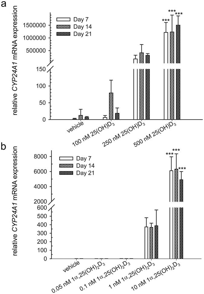
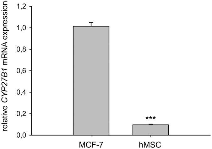
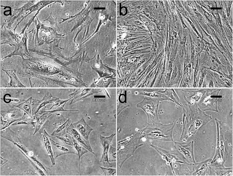
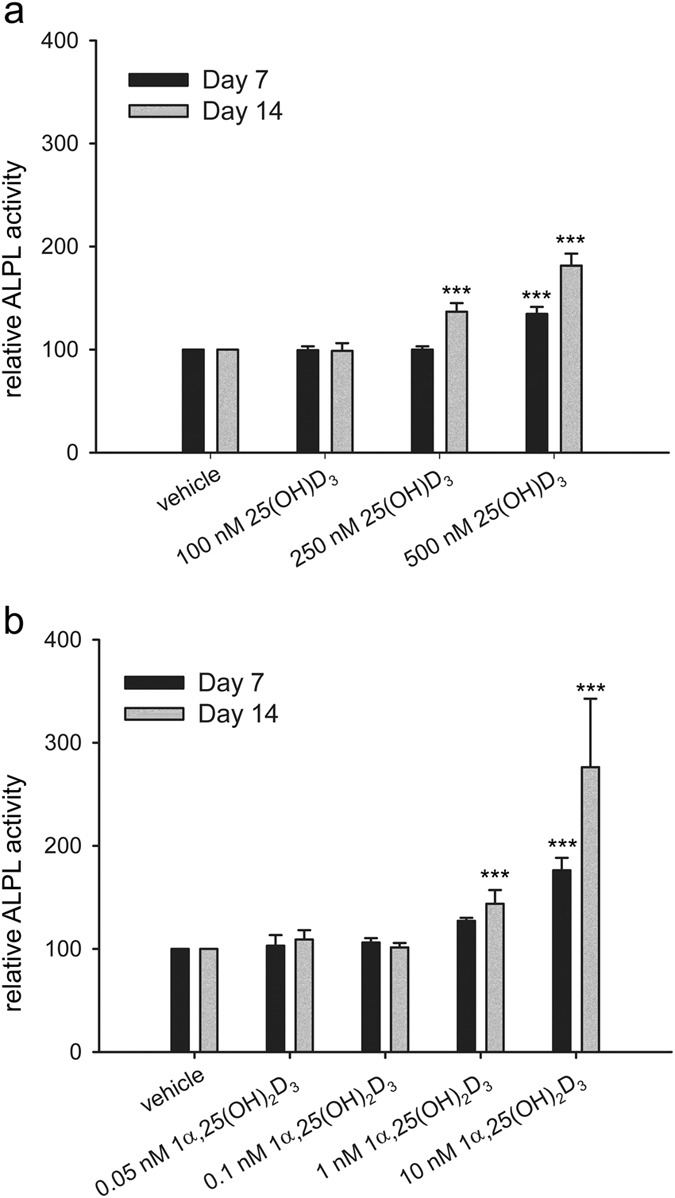
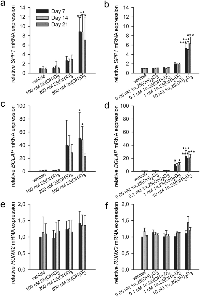
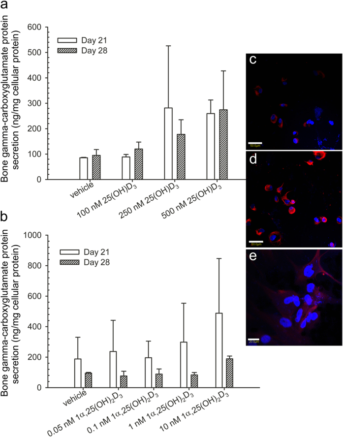
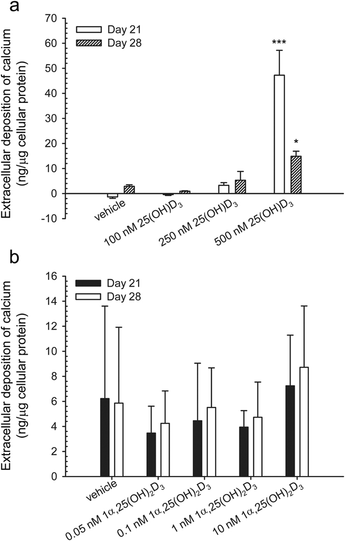
25-Hydroxyvitamin D3 [25(OH)D3] has recently been found to be an active hormone. Its biological actions are demonstrated in various cell types. 25(OH)D3 deficiency results in failure in bone formation and skeletal deformation. Here, we investigated the effect of 25(OH)D3 on osteogenic differentiation of human mesenchymal stem cells (hMSCs). We also studied the effect of 1α,25-dihydroxyvitamin D3 [1α,25-(OH)2D3], a metabolite of 25(OH)D3. One of the vitamin D responsive genes, 25(OH)D3-24-hydroxylase (cytochrome P450 family 24 subfamily A member 1) mRNA expression is up-regulated by 25(OH)D3 at 250-500 nM and by 1α,25-(OH)2D3 at 1-10 nM. 25(OH)D3 and 1α,25-(OH)2D3 at a time-dependent manner alter cell morphology towards osteoblast-associated characteristics. The osteogenic markers, alkaline phosphatase, secreted phosphoprotein 1 (osteopontin), and bone gamma-carboxyglutamate protein (osteocalcin) are increased by 25(OH)D3 and 1α,25-(OH)2D3 in a dose-dependent manner. Finally, mineralisation is significantly increased by 25(OH)D3 but not by 1α,25-(OH)2D3. Moreover, we found that hMSCs express very low level of 25(OH)D3-1α-hydroxylase (cytochrome P450 family 27 subfamily B member 1), and there is no detectable 1α,25-(OH)2D3 product. Taken together, our findings provide evidence that 25(OH)D3 at 250-500 nM can induce osteogenic differentiation and that 25(OH)D3 has great potential for cell-based bone tissue engineering.
Conflict of interest statement
The authors declare no competing financial interests.
Figures
Similar articles
-
TAp63γ and ΔNp63β promote osteoblastic differentiation of human mesenchymal stem cells: regulation by vitamin D3 Metabolites.PLoS One. 2015 Apr 7;10(4):e0123642. doi: 10.1371/journal.pone.0123642. eCollection 2015. PLoS One. 2015. PMID: 25849854 Free PMC article.
-
24R,25-dihydroxyvitamin D3 promotes the osteoblastic differentiation of human mesenchymal stem cells.Mol Endocrinol. 2014 May;28(5):644-58. doi: 10.1210/me.2013-1241. Epub 2014 Mar 5. Mol Endocrinol. 2014. PMID: 24597546 Free PMC article.
-
25-Hydroxy- and 1α,25-Dihydroxycholecalciferol Have Greater Potencies than 25-Hydroxy- and 1α,25-Dihydroxyergocalciferol in Modulating Cultured Human and Mouse Osteoblast Activities.PLoS One. 2016 Nov 28;11(11):e0165462. doi: 10.1371/journal.pone.0165462. eCollection 2016. PLoS One. 2016. PMID: 27893751 Free PMC article.
-
The renal function of 25-hydroxyvitamin D3-1alpha-hydroxylase.Mol Cell Endocrinol. 1999 May 25;151(1-2):213-20. doi: 10.1016/s0303-7207(99)00039-8. Mol Cell Endocrinol. 1999. PMID: 10411336 Review.
-
1alpha(OH)D3 One-alpha-hydroxy-cholecalciferol--an active vitamin D analog. Clinical studies on prophylaxis and treatment of secondary hyperparathyroidism in uremic patients on chronic dialysis.Dan Med Bull. 2008 Nov;55(4):186-210. Dan Med Bull. 2008. PMID: 19232159 Review.
Cited by
-
Identification of the genetic central dogma in osteogenic differentiation of MSCs by osteoinductive medium from transcriptional data sets.Chronic Dis Transl Med. 2022 May 31;8(3):218-228. doi: 10.1002/cdt3.26. eCollection 2022 Sep. Chronic Dis Transl Med. 2022. PMID: 36161200 Free PMC article.
-
Vitamin D sterols increase FGF23 expression by stimulating osteoblast and osteocyte maturation in CKD bone.Bone. 2019 Oct;127:626-634. doi: 10.1016/j.bone.2019.07.026. Epub 2019 Aug 1. Bone. 2019. PMID: 31377240 Free PMC article.
-
Burst, Short, and Sustained Vitamin D3 Applications Differentially Affect Osteogenic Differentiation of Human Adipose Stem Cells.Int J Mol Sci. 2020 Apr 30;21(9):3202. doi: 10.3390/ijms21093202. Int J Mol Sci. 2020. PMID: 32366057 Free PMC article.
-
Osteogenic and Angiogenic Synergy of Human Adipose Stem Cells and Human Umbilical Vein Endothelial Cells Cocultured in a Modified Perfusion Bioreactor.Organogenesis. 2021 Oct 2;17(3-4):56-71. doi: 10.1080/15476278.2021.1954769. Epub 2021 Jul 29. Organogenesis. 2021. PMID: 34323661 Free PMC article.
-
Phytochemical Compounds Involved in the Bone Regeneration Process and Their Innovative Administration: A Systematic Review.Plants (Basel). 2023 May 22;12(10):2055. doi: 10.3390/plants12102055. Plants (Basel). 2023. PMID: 37653972 Free PMC article. Review.
References
-
- DeLuca H. F. Overview of general physiologic features and functions of vitamin D. Am. J. Clin. Nutr. 80, 1689S–1696S (2004). - PubMed
-
- van Driel M. et al.. Evidence for auto/paracrine actions of vitamin D in bone: 1alpha-hydroxylase expression and activity in human bone cells. FASEB. J. 20, 2417–2419 (2006). - PubMed
-
- Holick M. F. Vitamin D deficiency. N. Engl. J. Med. 357, 266–281 (2007). - PubMed
-
- Guardia G. et al.. Prevalence of vitamin D depletion among subjects seeking advice on osteoporosis: a five-year cross-sectional study with public health implications. Osteoporos. Int. 19, 13–19 (2008). - PubMed
Publication types
MeSH terms
Substances
LinkOut - more resources
Full Text Sources
Other Literature Sources
Research Materials
Miscellaneous
