. 2017 Feb 17:7:43012.
doi: 10.1038/srep43012.

High-resolution 3D imaging of whole organ after clearing: taking a new look at the zebrafish testis

Affiliations

High-resolution 3D imaging of whole organ after clearing: taking a new look at the zebrafish testis

Maxence Frétaud et al. Sci Rep. .

Abstract

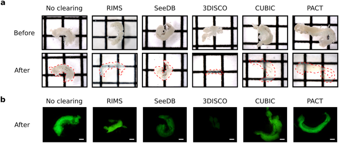
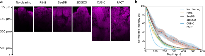
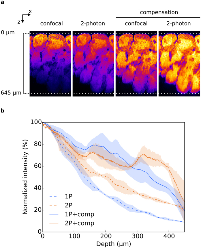
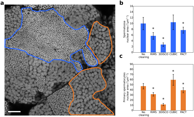
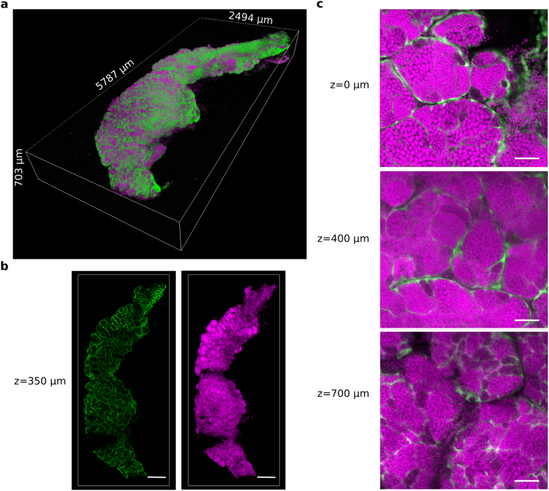
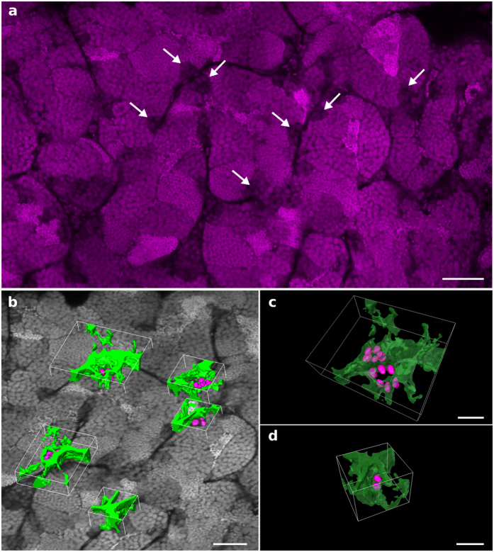
Zebrafish testis has become a powerful model for reproductive biology of teleostean fishes and other vertebrates and encompasses multiple applications in applied and basic research. Many studies have focused on 2D images, which is time consuming and implies extrapolation of results. Three-dimensional imaging of whole organs recently became an important challenge to better understand their architecture and allow cell enumeration. Several protocols have thus been developed to enhance sample transparency, a limiting step for imaging large biological samples. However, none of these methods has been applied to the zebrafish testis. We tested five clearing protocols to determine if some of them could be applied with only small modifications to the testis. We compared clearing efficiency at both macroscopic and microscopic levels. CUBIC and PACT were suitable for an efficient transparency, an optimal optical penetration, the GFP fluorescence preservation and avoiding meaningful tissue deformation. Finally, we succeeded in whole testis 3D capture at a cellular resolution with both CUBIC and PACT, which will be valuable in a standard workflow to investigate the 3D architecture of the testis and its cellular content. This paves the way for further development of high content phenotyping studies in several fields including development, genetic or toxicology.


Conflict of interest statement

The authors declare no competing financial interests.

Figures

Figure 1
Figure 1. Comparison of the transparency and the GFP fluorescence of testes treated with different clearing methods.
Testes were dissected from the zebrafish transgenic line Tg(gsdf:GFP) and cleared with RIMS, SeeDB, 3DISCO, CUBIC or PACT protocols. Testes were incubated in the refractive index matching solution of the last step of each protocol and imaged within 1 day. (a) Brightfield images of testes before and after clearing with the indicated methods. Transparency is assessed by the visualization of black lines situated underneath each sample. Dotted red line indicates the edge of testes after clearing. Square = 1.6 mm × 1.6 mm. (b) GFP fluorescence of cleared and non-cleared testes. The different clearing protocols used are indicated. Scale bar: 500 μm.
Figure 2
Figure 2. Comparison of the fluorescence recovery in depth from testes treated with different clearing protocols.
Two-photon imaging of testes cleared with RIMS, SeeDB, 3DISCO, CUBIC or PACT protocols. (a) XZ planes of testes. Nuclei were stained with propidium iodide (in magenta). Laser intensity was set in order to be next to saturation at the beginning of the stack and no depth compensation was used. No brightness and contrast enhancement was applied. (b) Quantification of fluorescence intensity. Mean fluorescence intensity was normalized and plotted against the imaging depth. Mean ± SEM of 4–12 ROI acquired on 2–3 different testes.
Figure 3
Figure 3. Comparison of the spatial resolution of images acquired from testes treated with different clearing protocols.
Two-photon imaging of testes cleared with RIMS, SeeDB, 3DISCO, CUBIC or PACT protocols. XY planes of testes at three different imaging depths: 15 μm, 150 μm and 300 μm. Nuclei were stained with propidium iodide (in magenta). Laser intensity was set in order to be next to saturation at the beginning of the stack and no depth compensation was used. Brightness and contrast has been modified to assess spatial resolution. Images were acquired at a scanning speed of 400 Hz and at a resolution of 1024 × 1024 pixels with two lines average. N/A: Not available because there is no tissue at this depth. Scale bar: 50 μm.
Figure 4
Figure 4. Comparison of confocal and 2-photon imaging on CUBIC cleared testes.
(a) XZ planes of testis treated with CUBIC and acquired either by confocal or 2-photon microscopy, with or without laser compensation. No brightness and contrast enhancement was applied. Nuclei were stained with propidium iodide and pseudocolored. (b) Fluorescence intensity quantification. Mean fluorescence intensity was normalized and plotted against the imaging depth. All data are mean ± SEM of 3 ROI acquired on 1 testis.
Figure 5
Figure 5. Comparison of the nuclear size of testes treated with different clearing protocols.
(a) Optical section of a non-cleared testis. Nuclei were stained with propidium iodide. Nuclei of germ cells at different stages of differentiation are packed in distinct domains. A spermatozoa domain is delineated in blue and a primary spermatocyte domain is delineated in orange. For each clearing condition (RIMS, 3DISCO, CUBIC and PACT), the nuclear area of 55 spermatozoa and 55 primary spermatocytes was measured, spreading across 11 optical sections from two testes. (b) Nuclear area of spermatozoa. (c) Nuclear area of primary spermatocytes. All data are indicated as mean ± SD. *Indicates a significant difference as compared with PBS (p < 0.0001) using a one way ANOVA with Dunnet’s post-hoc test for multiple comparison. Scale bar: 25 μm.
Figure 6
Figure 6. A 3D reconstruction of a whole zebrafish testis cleared with CUBIC.
A testis was dissected from the zebrafish transgenic line Tg(gsdf:GFP) and cleared with the CUBIC protocol. All nuclei are in magenta (propidum iodide). The somatic Sertoli cells are in green (endogenous GFP fluorescence). (a) 3D rendering of the whole CUBIC-cleared testis. (b) 2D optical sections of the testis at 350 μm in depth. (c) Magnified view of 2D optical sections at 0 μm, 400 μm and 700 μm in depth. Images were acquired in 8 bits at a scanning speed of 600 Hz and at a resolution of 512 × 512 pixels with two lines average. Scale bars: 500 μm (b) and 50 μm (c).
Figure 7
Figure 7. PACT 3D imaging of a zebrafish testis to study the architecture of germinal niches.
The testis dissected from a Tg(gsdf:GFP) transgenic line was stained with propidium iodide and cleared with the PACT protocol. Imaging and 3D reconstruction of the whole testis were performed as shown in Supplementary Fig. S5 and Supplementary Videos S6 and S7. (a) A 2D optical section at 237 μm in depth. Nuclei of undifferentiated spermatogonia are identified through the testis by their larger volume and low fluorescence intensity (arrows). (b) Examples of 3D surface reconstructions of germinal niches containing clustered undifferentiated spermatogonia. (c) High magnification view of two nearby niches located in adjacent seminiferous tubules and displaying six nuclei each. (d) High magnification view of a niche displaying two nuclei of undifferentiated spermatogonia. Nuclei are in grayscale or magenta. Surface reconstructed Sertoli cells and nuclei of undifferentiated spermatogonia are in green and magenta, respectively. Scale bars: 50 μm (a,b) and 20 μm (c).

References

    1. Hwang W. Y. et al.. Efficient genome editing in zebrafish using a CRISPR-Cas system. Nat. Biotechnol. 31, 227–229 (2013). - PMC - PubMed
    1. Hwang W. Y. et al.. Heritable and Precise Zebrafish Genome Editing Using a CRISPR-Cas System. PLoS One 8, e68708 (2013). - PMC - PubMed
    1. Varshney G. K. et al.. High-throughput gene targeting and phenotyping in zebrafish using CRISPR/Cas9. Genome Res. 25, 1030–1042 (2015). - PMC - PubMed
    1. Richardson D. S. & Lichtman J. W. Clarifying Tissue Clearing. Cell 162, 246–257 (2015). - PMC - PubMed
    1. Hama H. et al.. Scale: a chemical approach for fluorescence imaging and reconstruction of transparent mouse brain. Nat. Neurosci. 14, 1481–1488 (2011). - PubMed

Publication types

LinkOut - more resources